
Page 1623 - Williams Hematology ( PDFDrive )
P. 1623
1598 Part XI: Malignant Lymphoid Diseases Chapter 96: Pathology of Lymphomas 1599
The MYC gene most commonly is translocated to the IGH gene on chro-
mosome 14, resulting in t(8;14)(q24;q32), but it also can involve the
light-chain genes on chromosomes 2p12 (κ) and 22q11 (λ). A diagnosis
of Burkitt lymphoma can be suggested based on morphologic examina-
tion alone but should be supported by immunophenotypic data (posi-
tive for CD20, CD10, and BCL6; negative or focally weakly positive for
BCL2; growth fraction near 100 percent as determined by Ki67 stain)
and confirmed by molecular testing for MYC translocations whenever
possible.
Gene-expression studies have shown that Burkitt lymphoma has a
consistent gene-expression signature, but that there is not always cor-
relation between the diagnosis based on gene-expression profiling and
the diagnosis based on standard diagnostic testing. 53,54 To reflect this,
the 2008 WHO classification recognizes a provisional entity of B-cell
lymphoma, unclassifiable, with features intermediate between DLBCL
14
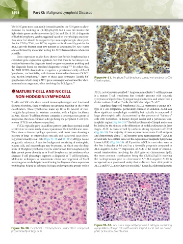
and Burkitt lymphoma. Many of these cases represent “double-hit” Figure 96–31. Peripheral T-cell lymphoma stained with antibody to CD3
lymphomas, which carry a MYC gene rearrangement and another chro- (T-cell marker).
mosomal rearrangement, often involving the BCL2 gene. 48
MATURE T-CELL AND NK CELL PTCL, not otherwise specified. Angioimmunoblastic T-cell lymphoma
55
NON-HODGKIN LYMPHOMAS is a mature T-cell lymphoma that typically presents with systemic
symptoms and polyclonal hypergammaglobulinemia, and arises from a
T cells and NK cells share several immunophenotypic and functional distinct subset of helper T cells, the follicular helper T cell. 56
features; therefore, these neoplasms are grouped together in the WHO Anaplastic large cell lymphoma (ALCL) represents a unique sub-
classification. These lymphomas make up 10 to 15 percent of non- type of T-cell lymphoma, particularly common in children. ALCL can
Hodgkin lymphomas in Western countries, with a higher incidence show significant morphologic variability but typically is composed of
in Asia. Mature T-cell lymphomas comprise a heterogeneous group of large pleomorphic cells characterized by the presence of “hallmark”
neoplasms, the most common subtype being the peripheral T-cell lym- cells with horseshoe- or kidney-shaped nuclei and a perinuclear eos-
phoma (PTCL) not otherwise specified. inophilic region (Fig. 96–32). Partial involvement of lymph nodes can
57
PTCLs typically grow in a diffuse pattern that effaces normal nodal be limited to the sinuses, with obliteration of nodal architecture in later
architecture or, more rarely, show expansion of the interfollicular areas. stages. ALCL is characterized by uniform, strong expression of CD30
They show a diverse cytologic spectrum, with most cases showing a (Fig. 96–33). The majority of cases express one or more T-cell antigens
mixture of large- to intermediate-size cells and occasional cases show- and demonstrate clonal T-cell receptor gene rearrangement. ALCL is
57
ing predominantly small cells (Figs. 96–30 and 96–31). Cell type has no divided into two entities based on the expression of anaplastic lymphoma
prognostic relevance. A reactive background consisting of eosinophils, kinase (ALK) (Fig. 96–34). ALK-positive ALCL is most often seen in
plasma cells, and macrophages may be present, in which case the diag- the first 3 decades of life and has a favorable prognosis compared to
nosis of Hodgkin lymphoma may be entertained. Immunophenotypic ALK-negative ALCL. 58,59 Expression of ALK is the result of chromo-
data cannot prove clonality as in B-cell lymphomas, but evidence of an somal translocations involving the ALK gene on chromosome 2p23,
aberrant T-cell phenotype supports a diagnosis of T-cell lymphoma. the most common translocation being the t(2;5)(p23;q35) involving
Molecular techniques to demonstrate clonal rearrangement of T-cell the nucleophosmin gene on chromosome 5. ALK-negative ALCL is
60
receptor genes can be helpful in confirming the diagnosis. Gene-expression recognized as a provisional entity that is distinct from ALK-positive
profiling has helped to delineate biologic and prognostic groups within ALCL and PTCL, not otherwise specified. Recently, additional genetic
61
Figure 96–32. Anaplastic large cell lymphoma, T-cell type, containing
Figure 96–30. Peripheral T-cell lymphoma, unspecified, composed a population of large cells with wreath-shaped nuclei and an eosino-
predominantly of large cells. philic perinuclear accentuation.
Kaushansky_chapter 96_p1587-1602.indd 1598 9/18/15 6:08 PM