
Page 1622 - Williams Hematology ( PDFDrive )
P. 1622
1596 Part XI: Malignant Lymphoid Diseases Chapter 96: Pathology of Lymphomas 1597
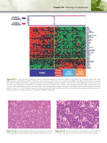
Figure 96–27. Gene-expression profiling of primary mediastinal large B-cell lymphoma (PMBCL), contrasting the expression profile with nodal
diffuse large B-cell lymphomas (DLBCL). This figure shows numerous genes that are overexpressed in PMBCL (red). Many of these genes are shared
with classical Hodgkin lymphoma, suggesting a biologic overlap between these two diseases. Cases listed as “Other Mediastinal” refer to those cases
of DLBCL with mediastinal involvement, but not felt to be typical of PMBCL. This is borne out by the gene-expression data, showing that these
cases are more closely related to DLBCL rather than PMBCL. ABC, activated B cell; GCB, germinal center B cell. (Reproduced with permission from Rosenwald A,
Wright G, Leroy K, Yu X, et al: Molecular siagnosis of primary mediastinal B cell lymphoma identifies a clinically favorable subgroup of diffuse large B cell
lymphoma related to Hodgkin Lymphoma. J Exp Med 15;198(6):851–862, 2003.)
Figure 96–28. Burkitt lymphoma with starry-sky appearance, imparted Figure 96–29. Burkitt lymphoma, characterized by a diffuse infiltrate
by macrophages that have engulfed apoptotic debris of dying tumor cells. of medium-size cells with small nucleoli and a high mitotic activity.
Kaushansky_chapter 96_p1587-1602.indd 1597 9/18/15 6:07 PM