
Page 1625 - Williams Hematology ( PDFDrive )
P. 1625
1600 Part XI: Malignant Lymphoid Diseases Chapter 96: Pathology of Lymphomas 1601
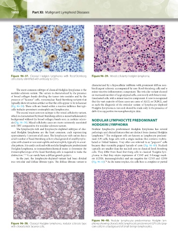
Figure 96–37. Classical Hodgkin lymphoma with Reed-Sternberg Figure 96–39. Mixed cellularity Hodgkin lymphoma.
cells clearly identified with antibody to CD15.
characterized by a hypocellular infiltrate with prominent diffuse non-
birefringent sclerosis accompanied by rare Reed-Sternberg cells and a
The most common subtype of classical Hodgkin lymphoma is the
nodular sclerosis variant. The variant is characterized by the presence minor reactive inflammatory component. The reticular variant showed
of broad collagen bands dividing the tumor into nodules and by the an increased number of large atypical cells, commonly with bizarre mul-
presence of “lacunar” cells, mononuclear Reed-Sternberg variants that tinucleated cells, with a minor reactive component. It now is recognized
typically show retraction artifact so that the cells appear to be in lacunae that the vast majority of these cases are cases of ALCL or DLBCL, and
(Fig. 96–38). These cells are found within a reactive infiltrate that typi- as such the diagnosis of the reticular variant of lymphocyte-depleted
cally includes prominent eosinophils and lymphocytes. Hodgkin lymphoma is rare and should be made only in the presence of
The second most common subtype is the mixed cellularity variant, definitive supportive immunophenotypic data.
which is characterized by Reed-Sternberg cells in a mixed inflammatory
background without the broad collagen bands seen in nodular sclero- NODULAR LYMPHOCYTE PREDOMINANT
sis (Fig. 96–39). Mixed cellularity cases are more commonly associated
with EBV compared to the nodular sclerosis variant. HODGKIN LYMPHOMA
The lymphocyte-rich and lymphocyte-depleted subtypes of clas- Nodular lymphocyte predominant Hodgkin lymphoma has several
sical Hodgkin lymphoma are the least common, each representing pathologic and clinical features that are distinct from classical Hodgkin
approximately 5 percent of all cases. The lymphocyte-rich variant has a lymphoma. The malignant cells are known as lymphocyte predomi-
73
small number of Reed-Sternberg cells in a background of small lympho- nant (LP) cells, large cells with a single nucleus that contains multilo-
cytes with absent or rare eosinophils and neutrophils, typically in a nod- bated or folded features. They often are referred to as “popcorn” cells
ular pattern. It is easily confused with nodular lymphocyte predominant because they resemble popped kernels of corn (Fig. 96–40). Nucleoli
Hodgkin lymphoma, so immunohistochemical stains to determine the typically are smaller than the nucleoli seen in classical Reed-Sternberg
immunophenotype of the Reed-Sternberg cells is required to make the cells. They differ from Reed-Sternberg cells in classical Hodgkin lym-
distinction. 71,72 It can rarely have a diffuse growth pattern. phoma in that they retain expression of CD45 and B-lineage mark-
In the past, the lymphocyte-depleted variant had been divided ers (CD20, immunoglobulin) and are negative for CD15 and CD30
into reticular and diffuse fibrosis types. The diffuse fibrosis variant is (Fig. 96–41). As the name implies, the cells have a complete or partial
74
Figure 96–40. Nodular lymphocyte predominance Hodgkin lym-
Figure 96–38. Classical Hodgkin lymphoma, nodular sclerosis type phoma showing characteristic lymphocyte predominant (LP) cells (pop-
with characteristic lacunar cells. corn cells) in a background of small benign lymphocytes.
Kaushansky_chapter 96_p1587-1602.indd 1600 9/18/15 6:08 PM