
Page 1620 - Williams Hematology ( PDFDrive )
P. 1620
1594 Part XI: Malignant Lymphoid Diseases Chapter 96: Pathology of Lymphomas 1595
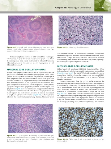
Figure 96–21. Lymph node involved by marginal zone B-cell lym- Figure 96–23. Diffuse large B-cell lymphoma.
phoma, in which the benign germinal centers and mantle zones are
surrounded by expanded pale marginal zones.
infection of the stomach. At early stages of development, many of these
39
lymphomas respond to treatment with antibiotics to eradicate H. pylori,
Follicular lymphoma in situ is an entity where BCL2-positive ger- whereas later changes, including cases with chromosomal transloca-
minal centers are present in an otherwise reactive lymph node. When tions activating genes involved in nuclear factor-κB (NF-κB) signaling,
40
it is distinguished from partial involvement by follicular lymphoma, lead to antigen-independent growth (Chap. 101).
follicular lymphoma in situ has a very low rate of progression to overt
follicular lymphoma. 36
DIFFUSE LARGE B-CELL LYMPHOMA
MARGINAL ZONE B-CELL LYMPHOMAS Diffuse large B-cell lymphoma (DLBCL) is characterized by a diffuse
Marginal zone lymphomas are characterized by a proliferation of small infiltrate of large B cells that can resemble centroblasts or immunoblasts
(Figs. 96–23 and 96–24). The 2008 WHO classification identifies several
lymphocytes, commonly with abundant pale cytoplasm (called mono- types of large B-cell lymphoma, the most common type being DLBCL
cytoid B cells) and plasmacytic features. The postulated cell of origin of not otherwise specified, which constitutes 25 to 30 percent of all non-
these lymphomas is the postgerminal center B cell of the marginal zone Hodgkin lymphomas.
at various anatomic sites. Marginal zone lymphomas can be divided into Gene-expression data show that DLBCL is a heterogeneous dis-
three distinct types based on site of presentation: (1) extranodal mar- ease consisting of at least three entities having distinct gene-expression
ginal zone lymphomas of mucosa-associated lymphoid tissue (MALT), profiles based on cell of origin: (1) cases with an expression profile sim-
37
(2) splenic marginal zone lymphomas, and (3) nodal marginal zone ilar to germinal center B cells (GCBs), (2) cases expressing genes typ-
lymphomas (Fig. 96–21). This classification is supported by distinctive ical of activated B cells (ABCs), and (3) cases with a different pattern
38
cytogenetic abnormalities in each entity. Extranodal lymphomas of the referred to as “unclassifiable” that are neither GCB-type nor ABC-type
MALT type are the most common and arise in mucosal sites subject (Fig. 96–25). Importantly, clinical differences were apparent, with
41
to longstanding chronic inflammation (Fig. 96–22), including chronic GCB-type cases having a significantly better prognosis compared to the
infection, the prototypical example being chronic Helicobacter pylori other two types, even when clinical prognostic markers are considered
(Chap. 98). Further studies confirmed these differences in the current
era of therapy (including anti-CD20 antibody therapy), and identified
Figure 96–22. Salivary gland involved by mucosa-associated lym-
phoid tissue (MALT) lymphoma, showing a diffuse infiltrate of small
lymphocytes with pale cytoplasm, infiltrating an enlarged salivary gland Figure 96–24. Diffuse large B-cell stained with antibody to CD20
duct (lymphoepithelial lesion). (B-cell marker).
Kaushansky_chapter 96_p1587-1602.indd 1595 9/18/15 6:07 PM