
Page 1621 - Williams Hematology ( PDFDrive )
P. 1621
1596 Part XI: Malignant Lymphoid Diseases Chapter 96: Pathology of Lymphomas 1597
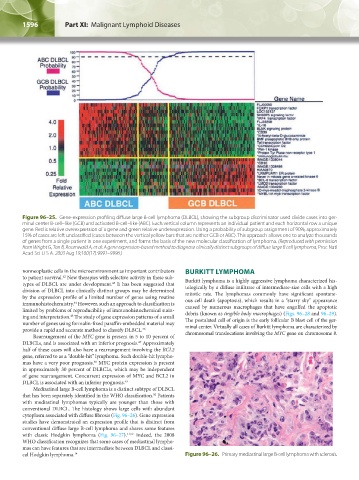
Figure 96–25. Gene-expression profiling diffuse large B-cell lymphoma (DLBCL), showing the subgroup discriminator used divide cases into ger-
minal center B-cell–like (GCB) and activated B-cell–like (ABC). Each vertical column represents an individual patient and each horizontal row a unique
gene. Red is relative overexpression of a gene and green relative underexpression. Using a probability of subgroup assignment of 90%, approximately
15% of cases are left unclassified (cases between the vertical yellow bars that are neither GCB or ABC). This approach allows one to analyze thousands
of genes from a single patient in one experiment, and forms the basis of the new molecular classification of lymphoma. (Reproduced with permission
from Wright G, Tan B, Rosenwald A, et al: A gene expression-based method to diagnose clinically distinct subgroups of diffuse large B cell lymphoma, Proc Natl
Acad Sci U S A. 2003 Aug 19;100(17):9991–9996.)
nonneoplastic cells in the microenvironment as important contributors BURKITT LYMPHOMA
to patient survival. New therapies with selective activity in these sub- Burkitt lymphoma is a highly aggressive lymphoma characterized his-
42
types of DLBCL are under development. It has been suggested that tologically by a diffuse infiltrate of intermediate-size cells with a high
43
division of DLBCL into clinically distinct groups may be determined mitotic rate. The lymphomas commonly have significant spontane-
by the expression profile of a limited number of genes using routine ous cell death (apoptosis), which results in a “starry sky” appearance
immunohistochemistry. However, such an approach to classification is caused by numerous macrophages that have engulfed the apoptotic
44
limited by problems of reproducibility of immunohistochemical stain- debris (known as tingible body macrophages) (Figs. 96–28 and 96–29).
ing and interpretation. The study of gene expression patterns of a small The postulated cell of origin is the early follicular B blast cell of the ger-
45
number of genes using formalin-fixed paraffin-embedded material may minal center. Virtually all cases of Burkitt lymphoma are characterized by
provide a rapid and accurate method to classify DLBCL. 46 chromosomal translocations involving the MYC gene on chromosome 8.
Rearrangement of the MYC gene is present in 5 to 10 percent of
DLBCLs, and is associated with an inferior prognosis. Approximately
47
half of these cases will also have a rearrangement involving the BCL2
gene, referred to as a “double-hit” lymphoma. Such double-hit lympho-
mas have a very poor prognosis. MYC protein expression is present
48
in approximately 30 percent of DLBCLs, which may be independent
of gene rearrangement. Concurrent expression of MYC and BCL2 in
DLBCL is associated with an inferior prognosis. 49
Mediastinal large B-cell lymphoma is a distinct subtype of DLBCL
that has been separately identified in the WHO classification. Patients
50
with mediastinal lymphomas typically are younger than those with
conventional DLBCL. The histology shows large cells with abundant
cytoplasm associated with diffuse fibrosis (Fig. 96–26). Gene expression
studies have demonstrated an expression profile that is distinct from
conventional diffuse large B-cell lymphoma and shares some features
with classic Hodgkin lymphoma (Fig. 96–27). 51,52 Indeed, the 2008
WHO classification recognizes that some cases of mediastinal lympho-
mas can have features that are intermediate between DLBCL and classi-
cal Hodgkin lymphoma. 14 Figure 96–26. Primary mediastinal large B-cell lymphoma with sclerosis.
Kaushansky_chapter 96_p1587-1602.indd 1596 9/18/15 6:07 PM