
Page 1633 - Williams Hematology ( PDFDrive )
P. 1633
1608 Part XI: Malignant Lymphoid Diseases Chapter 97: Hodgkin Lymphoma 1609
Detection of an unusual mass or swelling in the superficial, supra-
diaphragmatic lymph nodes (60 to 70 percent cervical and supraclavi-
cular, 15 to 20 percent axillary) is the most common presentation of cHL.
Only 15 to 20 percent of patients have subdiaphragmatic disease at
88
presentation. Lymphadenopathy is usually nontender and has a “rub-
bery” consistency. By inspection, a diffuse, puffy swelling rather than a
discrete mass may be apparent in the supraclavicular, infraclavicular, or
anterior chest wall regions. Infrequently, compression of the superior
vena cava will result in facial swelling and engorgement of the veins in
the neck and upper chest. Auscultation of the chest may reveal a pleural
effusion. Rarely, a significant pericardial effusion is present at diagnosis.
Palpation of the abdomen may reveal intraabdominal masses or hepa-
tosplenomegaly, although physical examination is relatively insensitive
for detection of these abnormalities.
Paraneoplastic Findings
A number of rare paraneoplastic syndromes have been described in A
cHL at the time of diagnosis. These include “vanishing bile duct syn-
drome” and idiopathic cholangitis with clinical jaundice, the neph-
rotic syndrome with anasarca, autoimmune hematologic disorders
(e.g., immune thrombocytopenia or hemolytic anemia), and neuro-
logic signs and symptoms. 89–91 Although parenchymal involvement
of the central nervous system or meningeal involvement is rare in
cHL, paraneoplastic syndromes include subacute cerebellar degener-
ation, myelopathy, progressive multifocal encephalopathy, and limbic
encephalitis. 91,92
RADIOGRAPHIC FEATURES
Intrathoracic disease is present at diagnosis in two-thirds of patients.
Mediastinal adenopathy is common in cHL, particularly in young
women with the nodular sclerosis subtype. Hilar adenopathy, pulmo-
93
nary parenchymal involvement, pleural effusions, pericardial effusions,
and chest wall masses may be appreciated by chest computed tomogra-
phy (CT); these are more common in the presence of extensive medi-
astinal disease. CT of the abdomen and pelvis is routinely employed in
the diagnostic evaluation of cHL. Although technologic advances have
greatly increased the resolution of this technique and the subsequent
detection of celiac, portal, splenic hilar, and mesenteric lymph nodes,
the correlation with histologic involvement of the spleen, historically B
determined by laparotomy staging, has been disappointing.
18
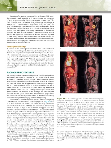
Whole-body F-fluorodeoxyglucose positron emission tomography Figure 97–5. Fluorodeoxyglucose positron emission tomography/
18
(FDG-PET) has become standard in the staging of cHL (Fig. 97–5). 94,95 computed tomography (FDG-PET/CT) imaging of classical Hodgkin
FDG-PET correlates well with CT evaluation and may demonstrate lymphoma. A. Coronal views of whole-body FDG-PET/CT imaging
performed before (May 31, 2011) and after (August 24, 2011) therapy
additional areas of disease, although this information uncommonly for stage IIB classical Hodgkin lymphoma with Adriamycin, bleomycin,
results in changes in stage or choice of initial therapy. 95,96 FDG-PET, vinblastine, and dacarbazine (ABVD) chemotherapy and involved field
however, is more sensitive to bone and hepatic disease and a diffuse radiotherapy. At the time of diagnosis, hypermetabolic areas of avid-
increase in signal can be seen at diagnosis in patients with neutrophilia. FDG uptake were evident in bilateral cervical, supraclavicular, medias-
FDG-PET imaging is superior to CT scanning in distinguishing active tinal, and hilar lymph nodes. Physiologic FDG uptake was visible in the
residual disease (increased glucose metabolism) from inactive residual colon and bladder. After completion of therapy, there was no abnormal
tissue, a major problem in assessing remission status after treatment, hypermetabolic activity in any of the original sites of disease, although
and has been incorporated in formal revised response guidelines. 94,95 physiologic FDG activity was seen in the cardiac blood cardiac blood
False-positive FDG-PET scans can be seen in the marrow during or at pool. B. Sagittal views of the same patient before (May 31, 2011) and
the end of treatment as a consequence of the chemotherapy effect or use after (August 24, 2011) therapy, showing resolution of hypermetabolism
in all involved nodal sites. The patient has remained in complete remis-
of hematopoietic colony-stimulating factors. In followup, false-positive sion since finishing therapy.
studies may be caused by thymic hyperplasia, granulomatous disease,
or infectious disorders. In addition to evaluation of residual masses,
FDG-PET has been incorporated in early response monitoring for those without concomitant abnormality on CT scan, usually require
risk stratification and, in clinical trials, to alter therapy. 96–102 The pre- tissue biopsy confirmation. Combined CT and FDG-PET technology
dictive accuracy of FDG-PET is dependent on expertise of the imag- is now standard for staging and posttreatment response evaluation and
ing staff and clinical correlation. In most situations, but particularly has resulted in improved anatomic definition of sites with increased
in FDG-PET, avid anatomic sites that were previously uninvolved or signal. 94–96
Kaushansky_chapter 97_p1603-1624.indd 1608 9/18/15 11:11 PM