
Page 1886 - Williams Hematology ( PDFDrive )
P. 1886
1860 Part XII: Hemostasis and Thrombosis Chapter 112: Platelet Morphology, Biochemistry, and Function 1861
One member of the integrin family, integrin α β , is virtually unique to Data from other integrin receptors identified a cell recognition
IIb 3
platelets (and their precursors, megakaryocytes), whereas the leucine-rich sequence composed of RGD in the ligand fibronectin, 819,820 and this
glycoproteins GPIb/IX and GPV appear to have highly restricted but same sequence is important in ligand binding to integrins α β and
V 3
not uniquely platelet expression patterns, including cytokine-activated α β . Fibrinogen contains one RGD sequence near the carboxy termi-
IIb 3
endothelial cells. 801,802 All of the other receptors are expressed more nus of each of the two Aα chains (amino acids 572 to 574) and another
821
widely on other cell types. at amino acids 95 to 97. In addition, the carboxyterminal 12 amino
acid region of each of the two γ chains (amino acids 400 to 411) contains
INTEGRINS a sequence that includes Lys-Gln-Ala-Gly-Asp-Val, which is the most
822–826
VWF contains
important in the binding of fibrinogen to platelets.
Integrin receptors are heterodimeric complexes composed of an α sub- an RGD sequence in its carboxyterminal domain and that region medi-
unit containing three or four divalent cation binding domains and a ates the binding to integrin α β . 809,810,812 Small, synthetic peptides con-
IIb 3
β subunit rich in disulfide bonds. Both subunits are transmembrane taining the RGD or γ-chain sequence inhibit the binding of fibrinogen
glycoproteins and are coded by different genes. There are at least 18 α to platelets, and these observations have been exploited to produce ther-
subunits and eight β subunits. 43,803,804 Three major families of integrin apeutic agents (tirofiban and eptifibatide) to inhibit platelet thrombus
receptors are recognized based on the β subunit: β , β , and β . Integrins formation (Chap. 134). Similarly, monoclonal antibodies that inhibit
827
3
2
1
are widely distributed on different cell types, and each integrin dem- binding of ligands to integrin α β have been developed and a mouse/
IIb 3
onstrates unique ligand-binding properties. Integrin receptors mediate human chimeric Fab fragment of one of them has been developed into a
interactions between cells and proteins or proteins on cells; they are also drug (abciximab) that is an effective antiplatelet agent.
involved in protein trafficking in cells. Integrin receptors can also trans- The binding of fibrinogen to integrin α β appears to be a mul-
IIb 3
duce messages from outside the cell to inside the cell, and from inside tistep process 808,828–833 : (1) the initial interaction is most likely via the
the cell to outside the cell. γ-chain carboxyterminal region(s) and divalent cation-dependent 823–826 ;
(2) subsequent interactions enhance the binding and internalization
Integrin α β (Also Termed GPIIb/IIIa, Fibrinogen Receptor, of the fibrinogen and render it irreversible, even when divalent cat-
834
IIb 3
and CD41/CD61) ions are removed ; (3) binding of fibrinogen induces changes in the
835
The integrin α β complex, a member of the β integrin receptor fam- receptor that can be recognized by antibodies (ligand-induced binding
3
IIb 3
ily, is the dominant platelet receptor, with 80,000 to 100,000 receptors sites [LIBSs]) 442,826 ; (4) binding of fibrinogen to integrin α β induces
IIb 3
present on the surface of a resting platelet (Fig. 112–11). 805–812 Another changes in fibrinogen (receptor-induced binding sites) that can be rec-
20,000 to 40,000 receptors are present inside platelets, primarily in ognized by antibodies and may involve exposure of the Aα chain Arg-
α-granule membranes, but also in dense bodies and membranes lining Gly-Asp-Phe sequence at amino acids 95 to 98 836,837 ; and (5) fibrinogen
the open canalicular system; these receptors are able to join the plasma binding induces receptor clustering. 251,838
membrane when platelets are activated and undergo the release reac- By electron microscopy, the receptors have a globular head of
tion. 813–815 On average, integrin α β receptors are less than 20 nm apart 8 × 12 nm and two 18-nm long tails representing the carboxyterminal
IIb 3
on the platelet surface and thus are among the most densely expressed regions of each subunit, including their hydrophobic transmembrane
adhesion/aggregation receptors present on any cell type. domains. 839,840 Crystallographic, electron microscopic, electron and
On resting platelets, integrin α β has low affinity for fibrinogen neutron scattering, and biochemical data from integrin α β and the
IIb 3
IIb 3
in solution, but when platelets are activated with ADP, epinephrine, related integrin α β receptor indicate that the unactivated receptors are
V 3
thrombin, or other agonists, integrin α β binds fibrinogen relatively in a bent conformation and that activation involves both extension of
IIb 3
strongly. 808,816 Activation induces changes in the integrin α β receptor the receptor head and a swing out motion in the β subunit. 149,827,841–853
3
IIb 3
itself that are responsible for the change in fibrinogen-binding affin- A three-dimensional reconstruction of integrin α β in a lipid bilayer
IIb 3
ity, but changes in the microenvironment surrounding integrin α β nano disc from negative-stain electron microscopy images supports a
IIb 3
may also be involved. The integrin α β receptors in α granules appear compact conformation of the inactive receptor, but unlike the crystal
IIb 3
to cycle to and from the plasma membrane. This recycling helps to structure of the ectodomain, the legs are not parallel and straight. 848
817
explain the ability of the integrin to take up fibrinogen from plasma and Integrin α β shares the same basic structural features of all integ-
IIb 3
transport it to α granules, where it is concentrated. 375,818 rin receptors (Table 112–4). 30,848 The α subunit, α , is a transmembrane
IIb
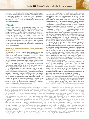
and actin-myosin contractile force. Ligand binding to the integrin is associated with a swing out motion of the integrin β hybrid domain from the
3
βA(I) domain (C), which results in both increased ligand affinity via alterations in the ADMIDAS (adjacent to metal ion-dependent adhesion site) and
MIDAS (metal ion-dependent adhesion site) regions of integrin β and greater leg separation. This conformational change may initiate outside-in
3
signaling. The ligated integrins may then cluster (not shown). The structure in panel (A) is based on the crystal structure of the ectodomain (PDB
250
3FCS) and the nuclear magnetic resonance (NMR) structure of the transmembrane and cytoplasmic domains (PDB 2K9J). The structure in (B) is
894
250
based on the same ectodomain crystal structure, but with extension at the genus of the subunits (PDB 3FCS), the NMR structures of the separated
transmembrane and cytoplasmic domains, and the structure of the complex between the β cytoplasmic domain and the talin F3 domain (PDB
894
3
827
2H7E). The structure in (C) is based on crystal structure of the liganded receptor (PDB 2VDN) headpiece, the extended structure of ectodomain
896
(PDB3FCS), and the monomeric transmembrane structures connected to unstructured cytosolic tails. B. Domain structure of structure of integrin
250
α β . The individual domains and the ligand binding pocket are identified in the model of the extended integrin. I-EGF, Integrin epidermal growth
IIb 3
factor; PSI, plexins, semaphorins, integrins. C. The integrin transmembrane complex. Selected views of the NMR structure of the α (red) and β (blue)
3
IIb
transmembrane complex. The left panel depicts contacts involved in the outer membrane clasp and the right panel depicts the contacts involved in
the inner membrane clasp. Note that after the integrin α helical region ends at V990, the next 5 residues (GFFKR) reenter the membrane; the two
IIb
aromatic F residues make hydrophobic contacts with β and α R995 makes a salt bridge with integrin β D723. (A, reproduced with permission from
3
IIb
3
Lau TL, Kim C, Ginsberg MH, et al: The structure of the integrin alphaIIbbeta3 transmembrane complex explains integrin transmembrane signalling. EMBO J
28(9):1351–1361, 2009. B, reproduced with permission from Zhu, J, et al: Structure of a complete integrin ectodomain in a physiologic resting state and activa-
tion and deactivation by applied forces. Mol Cell 32(6):849–861, 2008. C, reproduced with permission from Lau TL, Kim C, Ginsberg MH, et al: The structure of
the integrin alphaIIbbeta3 transmembrane complex explains integrin transmembrane signalling. EMBO J 28(9):1351–1361, 2009.)
Kaushansky_chapter 112_p1829-1914.indd 1861 17/09/15 3:29 pm