
Page 1965 - Williams Hematology ( PDFDrive )
P. 1965
1940 Part XII: Hemostasis and Thrombosis Chapter 113: Molecular Biology and Biochemistry of the Coagulation Factors 1941
cellular membranes under (reduced) flow are still complicated, which that “prime” the clotting system for a subsequent burst of thrombin
hampers their incorporation into a mathematical model. 370 generation. Experiments using tissue factor–activated whole blood
and cell-based systems have shown that platelets can be activated by
A Cell-Based Scheme of Coagulation thrombin that is generated by direct tissue factor–factor VIIa activation
The goal of coagulation is to produce a fibrin clot that seals the site of of factor Xa. 371–373 The small amounts of factor Va required for proth-
injury in the vessel wall. This process is initiated when tissue factor– rombinase assembly are likely provided by activated platelets, by factor
bearing cells are exposed to blood at the damaged site. Tissue factor is Xa activation, or potentially by noncoagulation proteases secreted by
anchored to cells via a transmembrane domain and acts as a receptor for the tissue factor–bearing cells. 335,374,375
plasma factor VII. Both trace amounts of factor VIIa as well as zymogen The small amounts of thrombin generated are capable of accom-
factor VII that is rapidly converted to factor VIIa by factor Xa and/or plishing the following: (1) activating platelets; (2) activating factor V;
autoactivation bind to tissue factor. Tissue factor is expressed around (3) activating factor VIII and dissociating factor VIII from VWF; and
vessels and in the epithelium, where it forms a “hemostatic envelope.” (4) activating factor XI (see Fig. 113–29). 366,372,373 The activity of the fac-
The tissue factor surrounding the vessels may already be in complex tor Xa formed by the tissue factor–factor VIIa complex will be mostly
with factor VIIa, even in the absence of an injury. 253 restricted to the tissue factor–bearing surface because free factor Xa that
The tissue factor–factor VIIa complex catalyzes two very impor- diffuses off the cell surface is rapidly inhibited by TFPI, AT, and/or the
tant reactions: (1) activation of factor X to factor Xa and (2) activation protein Z–ZPI complex. Factor IXa, on the other hand, will most likely
of factor IX to IXa. The initial factors Xa and IXa formed on tissue fac- act on activated platelets in close proximity to the tissue factor–bearing
tor–bearing cells may have distinct functions in initiating the process cell. This is because factor IXa can diffuse to adjacent cell surfaces as it is
371
of blood coagulation. When a vessel is damaged, the blood delivers not inhibited by TFPI and ZPI, while the rate of factor IXa inhibition by
platelets to the site of injury. These bind to extravascular matrix com- AT is much lower than that of factor Xa (see Table 113–4).
ponents to produce the primary hemostatic plug and become partially
activated in the process. The platelets are consequently localized in close The Role of Activated Platelets
proximity to active tissue factor–factor VIIa complexes. Platelets also play a major role in localizing clotting reactions to the
The factor Xa formed on the tissue factor–bearing cell interacts site of injury, as they adhere and aggregate at the same location where
with factor Va to form prothrombinase complexes that generate small tissue factor is exposed to blood. Platelet localization and activation are
amounts of thrombin (Fig. 113–29). Although this amount of thrombin mediated by VWF, thrombin, platelet receptors, and vessel wall com-
may not be sufficient to clot fibrinogen, it is sufficient to initiate events ponents such as collagen (Chap. 112). Once platelets are activated, the
cofactors Va and VIIIa are rapidly localized to the platelet membrane
X II surface (see Fig. 113–29). Cofactor binding is mediated in part by the
exposure of phosphatidylserine on the platelet membrane, a process
TFPI Xa VIII/VWF VIIIa + free VWF resulting from a flip-flop mechanism whereby phosphatidylserine on
Xa Vlla Vlla the inner leaflet of the membrane bilayer flips to the outer membrane
TF TF Xl
Va IIa leaflet. Endothelial cells, platelets, and leukocytes also generate pro-
376
V Platelet coagulant microvesicles that sustain thrombin generation. While the
Tissue factor–bearing cell V Xla procoagulant characteristics of microvesicles have been studied in
Va
detail in vitro, their relative contribution to coagulation in vivo is still
subject of debate.
TF Factor Xa generation is amplified on platelets by localization of
Vlla
IX X II factors IXa and XIa through specific binding sites, 377,378 and thrombin-
F mediated factor XI activation is enhanced by poly-P that is released
IXa by activated platelets (see Fig. 113–29). Once formed, factor Xa
379
IX Xa IIa associates with factor Va on the platelet surface to generate a burst of
XIa VIIIa
Va thrombin that is sufficient to clot fibrinogen and form a hemostatic
Activated platelet plug. Subsequently, thrombin-activated factor XIII crosslinks fibrin
and stabilizes the hemostatic plug, thereby rendering it impermeable.
Thrombin also activates TAFI, which helps to stabilize the fibrin clot.
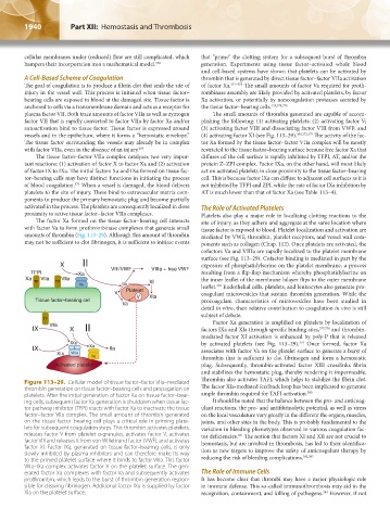
Figure 113–29. Cellular model of tissue factor–factor VIIa–mediated
thrombin generation on tissue factor–bearing cells and propagation on The factor XIa-mediated feedback loop has been implicated to generate
platelets. After the initial generation of factor Xa on tissue factor–bear- ample thrombin required for TAFI activation. 380
ing cells, subsequent factor Xa generation is shutdown when tissue fac- It should be noted that the balance between the pro- and anticoag-
tor pathway inhibitor (TFPI) reacts with factor Xa to inactivate the tissue ulant reactions, the pro- and antifibrinolytic potential, as well as stress
factor–factor VIIa complex. The small amount of thrombin generated on the local vasculature vary greatly in the different the organs, muscles,
on the tissue factor–bearing cell plays a critical role in priming plate- joints, and other sites in the body. This is probably fundamental to the
lets for subsequent coagulation steps. This thrombin activates platelets, variation in bleeding phenotypes observed in various coagulation fac-
releases factor V from platelet α-granules, activates factor V, activates tor deficiencies. The notion that factors XI and XII are not crucial to
381
factor VIII and releases it from von Willebrand factor (VWF), and activates hemostasis, but are involved in thrombosis, has led to their identifica-
factor XI. Factor IXa, generated on tissue factor–bearing cells, is only tion as new targets to improve the safety of anticoagulant therapy by
slowly inhibited by plasma inhibitors and can therefore make its way 382,383
to the primed platelet surface where it binds to factor VIIIa. This factor reducing the risk of bleeding complications.
VIIIa–IXa complex activates factor X on the platelet surface. The gen-
erated factor Xa complexes with factor Va and subsequently activates The Role of Immune Cells
prothrombin, which leads to the burst of thrombin generation respon- It has become clear that thrombi may have a major physiologic role
sible for cleaving fibrinogen. Additional factor IXa is supplied by factor in immune defense. This so-called immunothrombosis may aid in the
XIa on the platelet surface. recognition, containment, and killing of pathogens. However, if not
384
Kaushansky_chapter 113_p1915-1948.indd 1940 9/21/15 2:40 PM