
Page 1963 - Williams Hematology ( PDFDrive )
P. 1963
1938 Part XII: Hemostasis and Thrombosis Chapter 113: Molecular Biology and Biochemistry of the Coagulation Factors 1939
PROTEIN Z/PROTEIN Z–DEPENDENT PROTEASE the other Gla-containing coagulation proteins. There is an alternative
INHIBITOR exon that codes for a unique peptide of 22 amino acids in the prepro
leader sequence. The gene is transcribed into a 1.6-kb mRNA.
ZPI is a serine protease inhibitor (Mr ≈72,000; SERPINA10 in the sys- Several mutations and polymorphisms have been described for the
tematic nomenclature) that inhibits coagulation factors Xa and XIa. ZPI gene encoding ZPI, but the association between such gene variations
351
circulates in plasma at 60 nM with a half-life of 60 hours (see Table and risk of venous thrombosis has not been established with certainty. 352
343
113–1). The ZPI-dependent inhibition of factor Xa is enhanced in The human gene mutation database lists nine loss-of-function
79
the presence of protein Z. Protein Z is a vitamin K–dependent plasma mutations in the protein Z gene. The relationship between these muta-
GP (Mr ≈62,000) that circulates at 40 nM (see Table 113–1). In normal tions and disease is uncertain at best, but a relationship with ischemic
plasma, which has a molar excess of ZPI over protein Z, all protein Z stroke and recurrent fetal loss cannot be excluded. 353–355
circulates in complex with ZPI. 344
PATHWAYS OF HEMOSTASIS
Protein Structure
ZPI displays 25 to 30 percent homology with other serpins such as AT. Early Coagulation Schemes
Based on this homology, Tyr387 was predicted and confirmed as P1 res- With the accumulated knowledge of the biochemistry of hemophilia it
idue in the reactive center loop of ZPI and shown pivotal for inhibition was recognized in the 1960s that blood coagulation was regulated by a
345
of factor Xa. Unlike other serpins, the N-terminal region of ZPI con- sequential series of steps in which activation of one clotting factor led to
tains a very acidic domain. the activation of another, finally leading to a burst of thrombin genera-
356,357
Protein Z consists of a Gla domain, a hydrophobic region, and tion. Each clotting factor was thought to exist as a proenzyme that
two EGF-like domains. Even though the C-terminal region of protein could be converted to an active enzyme.
Z contains a domain that is homologous to the serine protease domains Since then the original waterfall reaction scheme of enzymes has
of the other Gla-containing proteins, it lacks the His and Ser active site been modified extensively. Factors V and VIII were identified as nonen-
residues characteristic for trypsin-like serine proteases. Thus, protein zymatic procofactors for factors Xa and IXa, respectively, and the subse-
346
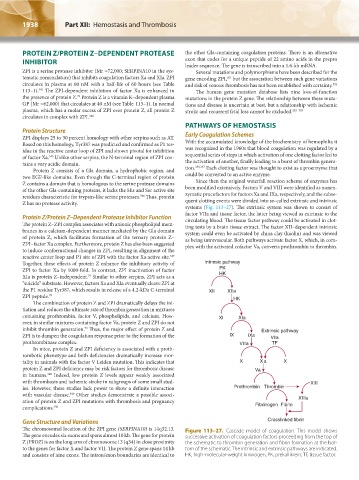
Z has no protease activity. quent clotting events were divided into so-called extrinsic and intrinsic
systems (Fig. 113–27). The extrinsic system was shown to consist of
factor VIIa and tissue factor, the latter being viewed as extrinsic to the
Protein Z/Protein Z–Dependent Protease Inhibitor Function circulating blood. The tissue factor pathway could be activated in clot-
The protein Z–ZPI complex associates with anionic phospholipid mem- ting tests by a brain tissue extract. The factor XII–dependent intrinsic
branes in a calcium-dependent manner mediated by the Gla domain system could even be activated by china clay (kaolin) and was viewed
of protein Z, which facilitates formation of the ternary protein Z– as being intravascular. Both pathways activate factor X, which, in com-
ZPI–factor Xa complex. Furthermore, protein Z has also been suggested plex with the activated cofactor Va, converts prothrombin to thrombin.
to induce conformational changes in ZPI, resulting in alignment of the
reactive center loop and P1 site of ZPI with the factor Xa active site.
347
Together, these effects of protein Z enhance the inhibitory activity of Intrinsic pathway
ZPI to factor Xa by 1000-fold. In contrast, ZPI inactivation of factor PK
79
XIa is protein Z–independent. Similar to other serpins, ZPI acts as a HK
“suicide” substrate. However, factors Xa and XIa eventually cleave ZPI at
the P1 residue Tyr387, which results in release of a 4.2-kDa C-terminal XII XIIa
ZPI peptide. 79 HK
The combination of protein Z and ZPI dramatically delays the ini-
tiation and reduces the ultimate rate of thrombin generation in mixtures
containing prothrombin, factor V, phospholipids, and calcium. How- XI XIa
ever, in similar mixtures containing factor Va, protein Z and ZPI do not
79
inhibit thrombin generation. Thus, the major effect of protein Z and Extrinsic pathway
ZPI is to dampen the coagulation response prior to the formation of the IX IXa Vlla
prothrombinase complex. Vllla TF
In mice, protein Z and ZPI deficiency is associated with a proth-
rombotic phenotype and both deficiencies dramatically increase mor-
tality in animals with the factor V Leiden mutation. This indicates that X X a X
protein Z and ZPI deficiency may be risk factors for thrombotic disease Va
348
in humans. Indeed, low protein Z levels appear weakly associated
with thrombosis and ischemic stroke in subgroups of some small stud- XIII
ies. However, these studies lack power to show a definite interaction Prothrombin Thrombin
with vascular disease. Other studies demonstrate a possible associ-
349
ation of protein Z and ZPI mutations with thrombosis and pregnancy XIIIa
complications. 350 Fibrinogen Fibrin
Gene Structure and Variations Crosslinked fibrin
The chromosomal location of the ZPI gene (SERPINA10) is 14q32.13. Figure 113–27. Cascade model of coagulation. This model shows
The gene encodes six exons and spans almost 10 kb. The gene for protein successive activation of coagulation factors proceeding from the top of
Z (PROZ) is on the long arm of chromosome 13 (q34) in close proximity the schematic to thrombin generation and fibrin formation at the bot-
to the genes for factor X and factor VII. The protein Z gene spans 14 kb tom of the schematic. The intrinsic and extrinsic pathways are indicated.
and consists of nine exons. The intron/exon boundaries are identical to HK, high-molecular-weight kininogen; PK, prekallikrein; TF, tissue factor.
Kaushansky_chapter 113_p1915-1948.indd 1938 9/21/15 2:40 PM