
Page 2133 - Williams Hematology ( PDFDrive )
P. 2133
2108 Part XII: Hemostasis and Thrombosis Chapter 122: The Vascular Purpuras 2109
A B
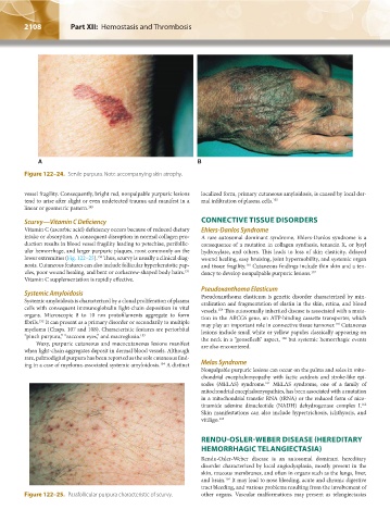
Figure 122–24. Senile purpura. Note accompanying skin atrophy.
vessel fragility. Consequently, bright red, nonpalpable purpuric lesions localized form, primary cutaneous amyloidosis, is caused by local der-
tend to arise after slight or even undetected trauma and manifest in a mal infiltration of plasma cells. 155
linear or geometric pattern. 149
Scurvy—Vitamin C Deficiency CONNECTIVE TISSUE DISORDERS
Vitamin C (ascorbic acid) deficiency occurs because of reduced dietary Ehlers-Danlos Syndrome
intake or absorption. A consequent disruption in normal collagen pro- A rare autosomal dominant syndrome, Ehlers-Danlos syndrome is a
duction results in blood vessel fragility leading to petechiae, perifollic- consequence of a mutation in collagen synthesis, tenascin X, or lysyl
ular hemorrhage, and larger purpuric plaques, most commonly on the hydroxylase, and others. This leads to loss of skin elasticity, delayed
lower extremities (Fig. 122–25). Thus, scurvy is usually a clinical diag- wound healing, easy bruising, joint hypermobility, and systemic organ
150
nosis. Cutaneous features can also include follicular hyperkeratotic pap- and tissue fragility. Cutaneous findings include thin skin and a ten-
156
ules, poor wound healing, and bent or corkscrew-shaped body hairs. dency to develop nonpalpable purpuric lesions. 157
151
Vitamin C supplementation is rapidly effective.
Pseudoxanthoma Elasticum
Systemic Amyloidosis Pseudoxanthoma elasticum is genetic disorder characterized by min-
Systemic amyloidosis is characterized by a clonal proliferation of plasma eralization and fragmentation of elastin in the skin, retina, and blood
cells with consequent immunoglobulin light-chain deposition in vital vessels. This autosomally inherited disease is associated with a muta-
158
organs. Microscopic 8 to 10 nm protofilaments aggregate to form tion in the ABCC6 gene, an ATP-binding cassette transporter, which
fibrils. It can present as a primary disorder or secondarily to multiple may play an important role in connective tissue turnover. Cutaneous
152
159
myeloma (Chaps. 107 and 108). Characteristic features are periorbital lesions include small white or yellow papules classically appearing on
“pinch purpura,” “raccoon eyes,” and macroglosia. 153 the neck in a “gooseflesh” aspect, but systemic hemorrhagic events
160
Waxy, purpuric cutaneous and mucocutaneous lesions manifest are also encountered.
when light-chain aggregates deposit in dermal blood vessels. Although
rare, palmodigital purpura has been reported as the sole cutaneous find-
ing in a case of myeloma-associated systemic amyloidosis. A distinct Melas Syndrome
154
Nonpalpable purpuric lesions can occur on the palms and soles in mito-
chondrial encephalomyopathy with lactic acidosis and stroke-like epi-
sodes (MELAS) syndrome. MELAS syndrome, one of a family of
161
mitochondrial encephalomyopathies, has been associated with a mutation
in a mitochondrial transfer RNA (tRNA) or the reduced form of nico-
tinamide adenine dinucleotide (NADH) dehydrogenase complex I.
162
Skin manifestations can also include hypertrichosis, ichthyosis, and
vitiligo. 163
RENDU-OSLER-WEBER DISEASE (HEREDITARY
HEMORRHAGIC TELANGIECTASIA)
Rendu-Osler-Weber disease is an autosomal dominant hereditary
disorder characterized by local angiodysplasia, mostly present in the
skin, mucous membranes, and often in organs such as the lungs, liver,
and brain. It may lead to nose bleeding, acute and chronic digestive
164
tract bleeding, and various problems resulting from the involvement of
Figure 122–25. Parafollicular purpura characteristic of scurvy. other organs. Vascular malformations may present as telangiectasias
Kaushansky_chapter 122_p2097-2112.indd 2108 9/18/15 10:30 AM