
Page 2131 - Williams Hematology ( PDFDrive )
P. 2131
2106 Part XII: Hemostasis and Thrombosis Chapter 122: The Vascular Purpuras 2107
Figure 122–20. Erythema multiforme. This hypersensitivity reaction,
usually to one of various drugs, characteristically presents with targetoid
lesions.
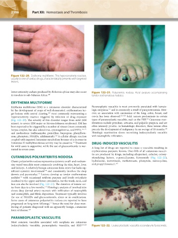
lower-extremity eschars produced by Rickettsia africae may also occur Figure 122–21. Polyarteritis nodosa. Acral purpura accompanying
in travelers to sub-Saharan Africa. 108 tender erythematous nodules.
ERYTHEMA MULTIFORME
Erythema multiforme (EM) is a cutaneous disorder characterized Paraneoplastic vasculitis is most commonly associated with hemato-
121
by the development of crops of well-demarcated, erythematous tar- logic neoplasia, and is commonly a result of paraproteinemia. How-
109
get lesions with central clearing, most commonly representing a ever, an association with carcinomas of the lung, colon, breast, and
hypersensitivity reaction triggered by infection or drug exposure cervix has been observed. 122–124 Solid tumors predominate in certain
125
(Fig. 122–20). The severity of this disorder ranges from mild (EM types of paraneoplastic vasculitis, such as the HSP. Cutaneous man-
minor), to severe (EM major or Stevens-Johnson syndrome). EM has ifestations include petechiae, urticaria, and palpable purpura, and are
been reported to be triggered by a number of viruses (most commonly often intensely pruritic. In hematologic disorders, these lesions often
126
herpes simplex, but also adenovirus, cytomegalovirus, and HIV), 110,111 precede the development of malignancy by an average of 10 months.
and medications (sulfonamides, penicillins, bupropion, phenylbuta- Histologic examination shows necrotizing leukocytoclastic vasculitis
112
zone, phenytoin, NSAIDs, adalimumab). A cellular allergic reaction with neutrophilic infiltration.
coupled with impaired histamine metabolism because of a decrease in
histamine-N-methyltransferase activity may be causative. Treatment DRUG-INDUCED VASCULITIS
113
for mild cases is supportive, while the use of glucocorticoids is war- A long list of drugs are reported to cause a vasculitis resulting in
ranted in severe cases.
erythematous purpuric lesions. One-fifth of all cutaneous vasculi-
tis are produced by drugs, including allopurinol, cefaclor, colony-
CUTANEOUS POLYARTERITIS NODOSA stimulating factors, d-penicillamine, furosemide (Fig. 122–22),
Classic polyarteritis nodosa represents a systemic small- and medium- hydralazine, isotretinoin, methotrexate, phenytoin, minocycline,
size vessel vasculitis most commonly involving the skin, heart, liver, and propylthiouracil. 127
and kidneys. A relatively benign cutaneous form exists that lacks sig-
nificant systemic involvement and consistently involves the deep
114
dermis and panniculus. Lesions develop as tender erythematous
115
nodules with occasional retiform purpura and livedo reticularis
116
localized to the upper and lower extremities, but the trunk, neck, and
face can also be involved (Fig. 122–21). The duration of lesions var-
ies from days to a few months. Histologic analysis of involved skin
115
shows deep dermal artery necrosis with infiltration of neutrophils
and eosinophils, and fibrin deposition. Treatment typically involves
the use of NSAIDs and glucocorticoids, alone or in combination.
Some cases of cutaneous polyarteritis nodosa are reported to have
progressed on long-term followup, hence the need for close mon-
117
itoring of patients diagnosed with an apparently benign, cutaneous
form of disease. 118
PARANEOPLASTIC VASCULITIS
Most common vasculitis associated with neoplasia are cutaneous
leukocytoclastic vasculitis, paraneoplastic vasculitis, and HSP. 119,120 Figure 122–22. Leukocytoclastic vasculitis secondary to furosemide.
Kaushansky_chapter 122_p2097-2112.indd 2106 9/18/15 10:30 AM