
Page 2130 - Williams Hematology ( PDFDrive )
P. 2130
2104 Part XII: Hemostasis and Thrombosis Chapter 122: The Vascular Purpuras 2105
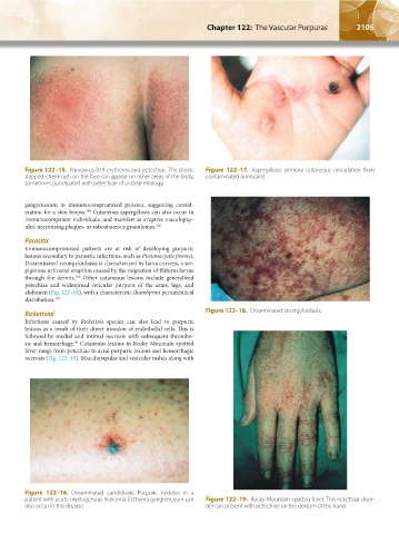
Figure 122–15. Parvovirus B19 erythema and petechiae. The classic Figure 122–17. Aspergillosis: primary cutaneous inoculation from
slapped-cheek rash on the face can appear on other areas of the body, contaminated armboard.
sometimes punctuated with petechiae of unclear etiology.
gangrenosum in immunocompromised patients, suggesting consid-
eration for a skin biopsy. Cutaneous aspergillosis can also occur in
104
immunocompetent individuals, and manifest as eruptive maculopap-
ules, necrotizing plaques, or subcutaneous granulomas. 105
Parasitic
Immunocompromised patients are at risk of developing purpuric
lesions secondary to parasitic infections, such as Pneumocystis jiroveci.
Disseminated strongyloidiasis is characterized by larva currens, a ser-
piginous urticarial eruption caused by the migration of filiform larvae
through the dermis. Other cutaneous lesions include generalized
106
petechiae and widespread reticular purpura of the arms, legs, and
abdomen (Fig. 122–18), with a characteristic thumbprint periumbilical
distribution. 107
Figure 122–18. Disseminated strongyloidiasis.
Rickettsial
Infections caused by Rickettsia species can also lead to purpuric
lesions as a result of their direct invasion of endothelial cells. This is
followed by medial and intimal necrosis with subsequent thrombo-
sis and hemorrhage. Cutaneous lesions in Rocky Mountain spotted
86
fever range from petechiae to acral purpuric lesions and hemorrhagic
necrosis (Fig. 122–19). Maculopapular and vesicular rashes along with
Figure 122–16. Disseminated candidiasis. Purpuric nodules in a
patient with acute myelogenous leukemia. Ecthyma gangrenosum can Figure 122–19. Rocky Mountain spotted fever. This rickettsial disor-
also occur in this disease. der can present with petechiae on the dorsum of the hand.
Kaushansky_chapter 122_p2097-2112.indd 2105 9/18/15 10:30 AM