
Page 2139 - Williams Hematology ( PDFDrive )
P. 2139
2114 Part XII: Hemostasis and Thrombosis Chapter 123: Hemophilia A and Hemophilia B 2115
activated the next zymogen until thrombin ultimately was produced. hemophilic males are normal, whereas all the daughters are obligatory
In this scheme, factors VIII and IX were considered to be proenzymes. carriers of the factor VIII defect. Sons of carriers have a 50 percent
Later, however, factor VIII, when activated by thrombin, was shown chance of being affected, whereas daughters of carriers have a 50 percent
not to be a proenzyme but rather an essential cofactor for factor IXa. chance of being carriers themselves.
The waterfall hypothesis has been modified so that the primary role of The factor VIII gene is very large, approximately 186 kb, with
the tissue factor–factor VII complex in the initiation of coagulation is approximately 9 kb of exons. The gene contains 26 exons and 25 introns.
16
emphasized (Chap. 113). 12 Based on the sequence of the factor VIII gene in normal individuals and
patients with hemophilia A, numerous specific mutations have been
ETIOLOGY AND PATHOGENESIS described 16,17 ; as of 2015, more than 2000 specific variants in the factor
17
Hemophilia A is a heterogeneous disorder resulting from defects in the VIII gene resulting in classic hemophilia have been described.
Hemophilia A can result from multiple alterations in the factor VIII
factor VIII gene that leads to absent or reduced circulating levels of func- gene. These include gene rearrangements; missense mutations, in which
tional factor VIII. The reduced activity can result from a decreased amount a single base substitution leads to an amino acid change in the molecule;
of factor VIII protein, the presence of a functionally abnormal protein, nonsense mutations, which result in a stop codon; abnormal splicing
or a combination of both. For factor VIII to be an effective cofactor for of the gene; deletions of all or portions of the gene; and insertions of
factor IXa, it must first be activated by thrombin, a reaction that results genetic elements. The genetic defects leading to hemophilia have been
18
in the formation of a heterotrimer composed of the A , A , A , C , and reviewed. 17
1
1
3
2
C domains of factor VIII in a complex with calcium (Chap. 113). One of the most common mutations, accounting for 40 to
13
2
Activated factor VIII (factor VIIIa) and activated factor IX (factor IXa) 50 percent of severe hemophilia A patients, is a unique “combined
associate on the surface of activated platelets, forming a functional factor gene inversion and crossing over” that disrupts the factor VIII gene. 19,20
X-activating complex (“tenase” or “Xase”). In the presence of factor VIIIa, Figures 123–2 and 123–3 schematically depict the factor VIII gene and
14
the rate of factor X activation by factor IXa is dramatically enhanced. That the mechanism of the “inversion–crossing over.” Within intron 22 are
21
hemophilia A and hemophilia B have similar clinical manifestations is not two other genes: (1) F8A(a ), which is transcribed in the 5′ direction,
1
surprising, because both factor VIIIa and factor IXa are required to form and (2) F8B, which is transcribed in the 3′ direction of the factor VIII
the Xase complex. The lack of either activated protein leads to a similar gene. The hatched boxes in Figure 123–3 show two other extragenic
lack of platelet surface Xase activity with subsequent decreased thrombin homologous sequences (a ,a ) 5′ to the F8A gene that lies within intron
2
3
generation. In patients with hemophilia, clot formation is delayed because 22 (a ). The presence of extragenic F8A sequences 5′ to the F8A gene
1
of the decreased thrombin generation. The clot that is formed is friable, within intron 22 is central to the inversion and translocation of part of
easily dislodged, and highly susceptible to fibrinolysis, all of which lead to the factor VIII gene from exon 1 to exon 22. The mechanism is homol-
excessive bleeding and poor wound healing. 15 ogous recombination between the F8A sequence that lies within intron
22 and one of the homologous extragenic sequences of the F8A gene
GENETICS 5′ to the factor VIII gene. During meiosis, crossing over of homolo-
Hemophilia A results when mutations occur in the factor VIII gene gous sequences occurs between the F8A gene lying within intron 22 and
located on the long arm of the X-chromosome (X-q28). The disease one of the extragenic homologous F8A sequences 5′ to intron 22. Thus,
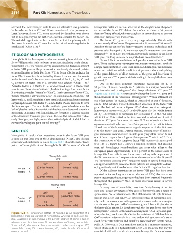
occurs almost exclusively in males. Figure 123–1 shows the inheritance the transcription of the complete factor VIII sequence is interrupted
pattern of hemophilia A and hemophilia B. All the sons of affected (Fig. 123–3). Figure 123–3 shows a common inversion and crossing
over, but homologous recombinations can occur with either of the
extragenic genes. Approximately 2 to 5 percent of the severe cases of
hemophilia A carry the intron 1 inversion resulting in the separation of
Hemophilic male the F8 promoter-exon 1 sequence from the remainder of the F8 gene.
22
h
X Y
The “inversion–crossing over” mutations result in severe hemophilia,
XX h XY and approximately 20 percent of these patients are susceptible to devel-
Normal X (Carrier female) (Normal male) oping antibody inhibitors that neutralize factor VIII coagulant function.
female X h Of the different insertions in the factor VIII gene that have been
XX XY reported, a few are long interspersed elements (LINEs) that are trans-
(Carrier female) (Normal male) poson sequences; that is, sequences that have been inserted frequently
throughout the genome. Most of these insertions result in severe
23
Normal male hemophilia.
XY In many cases of hemophilia, there is no family history of the dis-
ease, and at least 30 percent of the cases of hemophilia are a result of
h
XX h X Y spontaneous (de novo) mutations. Most of these occur at CpG dinucleo-
(Carrier female) (Hemophilic male)
23
Carrier X h tides in the factor VIII gene. De novo occurrences of hemophilia usu-
female X XX XY ally result from a mutation in the gamete of a normal male; for example,
(Normal female) (Normal male) a mutation in the germ cell of a maternal grandfather will give rise to
the hemophilia gene in his daughters such that his grandsons may have
18
hemophilia. Codons for the amino acid arginine (CGA [cytosine, gua-
Figure 123–1. Inheritance pattern of hemophilia. All daughters of a nine, adenine]) are frequently affected by mutations at CG doublets. A
hemophilic male are carriers of hemophilia, whereas all sons are nor- C→T transition often results in a stop codon with synthesis of a trun-
mal. Daughters of carriers have a 50 percent chance of being a carrier,
whereas sons of carriers have a 50 percent chance of having hemophilia. cated factor VIII molecule and usually is associated with severe hemo-
X, normal; X , abnormal X chromosome with the hemophilic gene; X Y, philia A. However, a G→A transition results in a missense mutation,
h
h
hemophilic male; XX, normal female; XX , carrier female; XY, normal which often leads to a dysfunctional factor VIII molecule that may be
h
male; Y, normal. associated with mild, moderate, or severe hemophilia. Some missense
Kaushansky_chapter 123_p2113-2132.indd 2114 9/21/15 4:35 PM