
Page 2142 - Williams Hematology ( PDFDrive )
P. 2142
2116 Part XII: Hemostasis and Thrombosis Chapter 123: Hemophilia A and Hemophilia B 2117
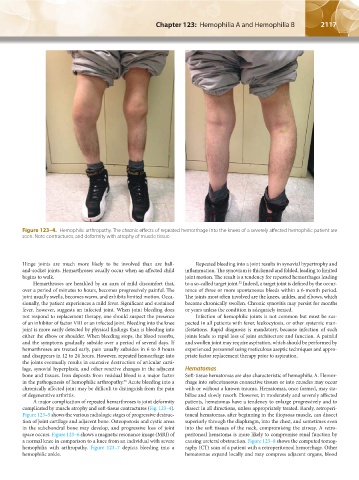
Figure 123–4. Hemophilic arthropathy. The chronic effects of repeated hemorrhage into the knees of a severely affected hemophilic patient are
seen. Note contractures, and deformity with atrophy of muscle tissue.
Hinge joints are much more likely to be involved than are ball- Repeated bleeding into a joint results in synovial hypertrophy and
and-socket joints. Hemarthroses usually occur when an affected child inflammation. The synovium is thickened and folded, leading to limited
begins to walk. joint motion. The result is a tendency for repeated hemorrhages leading
Hemarthroses are heralded by an aura of mild discomfort that, to a so-called target joint. Indeed, a target joint is defined by the occur-
32
over a period of minutes to hours, becomes progressively painful. The rence of three or more spontaneous bleeds within a 6-month period.
joint usually swells, becomes warm, and exhibits limited motion. Occa- The joints most often involved are the knees, ankles, and elbows, which
sionally, the patient experiences a mild fever. Significant and sustained become chronically swollen. Chronic synovitis may persist for months
fever, however, suggests an infected joint. When joint bleeding does or years unless the condition is adequately treated.
not respond to replacement therapy, one should suspect the presence Infection of hemophilic joints is not common but must be sus-
of an inhibitor of factor VIII or an infected joint. Bleeding into the knee pected in all patients with fever, leukocytosis, or other systemic man-
joint is more easily detected by physical findings than is bleeding into ifestations. Rapid diagnosis is mandatory, because infection of such
either the elbow or shoulder. When bleeding stops, the blood resorbs, joints leads to rapid loss of joint architecture and function. A painful
and the symptoms gradually subside over a period of several days. If and swollen joint may require aspiration, which should be performed by
hemarthroses are treated early, pain usually subsides in 6 to 8 hours experienced personnel using meticulous aseptic techniques and appro-
and disappears in 12 to 24 hours. However, repeated hemorrhage into priate factor replacement therapy prior to aspiration.
the joints eventually results in extensive destruction of articular carti-
lage, synovial hyperplasia, and other reactive changes in the adjacent Hematomas
bone and tissues. Iron deposits from residual blood is a major factor Soft-tissue hematomas are also characteristic of hemophilia A. Hemor-
in the pathogenesis of hemophilic arthropathy. Acute bleeding into a rhage into subcutaneous connective tissues or into muscles may occur
33
chronically affected joint may be difficult to distinguish from the pain with or without a known trauma. Hematomas, once formed, may sta-
of degenerative arthritis. bilize and slowly resorb. However, in moderately and severely affected
A major complication of repeated hemarthroses is joint deformity patients, hematomas have a tendency to enlarge progressively and to
complicated by muscle atrophy and soft-tissue contractures (Fig. 123–4). dissect in all directions, unless appropriately treated. Rarely, retroperi-
Figure 123–5 shows the various radiologic stages of progressive destruc- toneal hematomas, after beginning in the iliopsoas muscle, can dissect
tion of joint cartilage and adjacent bone. Osteoporosis and cystic areas superiorly through the diaphragm, into the chest, and sometimes even
in the subchondral bone may develop, and progressive loss of joint into the soft tissues of the neck, compromising the airway. A retro-
space occurs. Figure 123–6 shows a magnetic resonance image (MRI) of peritoneal hematoma is more likely to compromise renal function by
a normal knee in comparison to a knee from an individual with severe causing ureteral obstruction. Figure 123–8 shows the computed tomog-
hemophilia with arthropathy. Figure 123–7 depicts bleeding into a raphy (CT) scan of a patient with a retroperitoneal hemorrhage. Other
hemophilic ankle. hematomas expand locally and may compress adjacent organs, blood
Kaushansky_chapter 123_p2113-2132.indd 2117 9/21/15 4:35 PM