
Page 2141 - Williams Hematology ( PDFDrive )
P. 2141
2116 Part XII: Hemostasis and Thrombosis Chapter 123: Hemophilia A and Hemophilia B 2117
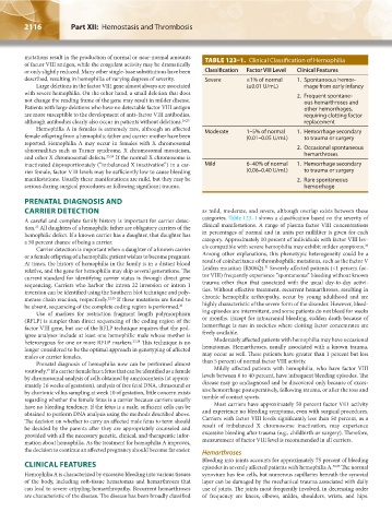
mutations result in the production of normal or near-normal amounts TABLE 123–1. Clinical Classification of Hemophilia
of factor VIII antigen, while the coagulant activity may be dramatically
or only slightly reduced. Many other single-base substitutions have been Classification Factor VIII Level Clinical Features
described, resulting in hemophilia of varying degrees of severity. Severe ≤1% of normal 1. Spontaneous hemor-
Large deletions in the factor VIII gene almost always are associated (≤0.01 U/mL) rhage from early infancy
with severe hemophilia. On the other hand, a small deletion that does 2. Frequent spontane-
not change the reading frame of the gene may result in milder disease. ous hemarthroses and
Patients with large deletions who have no detectable factor VIII antigen other hemorrhages,
are more susceptible to the development of anti–factor VIII antibodies, requiring clotting factor
although antibodies clearly also occur in patients without deletions. 16,23 replacement
Hemophilia A in females is extremely rare, although an affected Moderate 1–5% of normal 1. Hemorrhage secondary
female offspring from a hemophilic father and carrier mother have been (0.01–0.05 U/mL) to trauma or surgery
reported. Hemophilia A may occur in females with X chromosomal
abnormalities such as Turner syndrome, X chromosomal mosaicism, 2. Occasional spontaneous
and other X chromosomal defects. 23,24 If the normal X chromosome is hemarthroses
inactivated disproportionately (“imbalanced X inactivation”) in a car- Mild 6–40% of normal 1. Hemorrhage secondary
rier female, factor VIII levels may be sufficiently low to cause bleeding (0.06–0.40 U/mL) to trauma or surgery
manifestations. Usually these manifestations are mild, but they may be 2. Rare spontaneous
serious during surgical procedures or following significant trauma. hemorrhage
PRENATAL DIAGNOSIS AND
CARRIER DETECTION as mild, moderate, and severe, although overlap exists between these
A careful and complete family history is important for carrier detec- categories. Table 123–1 shows a classification based on the severity of
25
tion. All daughters of a hemophilic father are obligatory carriers of the clinical manifestations. A range of plasma factor VIII concentrations
hemophilic defect. If a known carrier has a daughter, that daughter has in percentages of normal and in units per milliliter is given for each
a 50 percent chance of being a carrier. category. Approximately 10 percent of individuals with factor VIII lev-
30
Carrier detection is important when a daughter of a known carrier els compatible with severe hemophilia may exhibit milder symptoms.
or a female offspring of a hemophilic patient wishes to become pregnant. Among other explanations, this phenotypic heterogeneity could be a
At times, the history of hemophilia in the family is in a distant blood result of coinheritance of thrombophilic mutations, such as the factor V
31
relative, and the gene for hemophilia may skip several generations. The Leiden mutation (R506Q). Severely affected patients (<1 percent fac-
current standard for identifying carrier status is through direct gene tor VIII) frequently experience “spontaneous” bleeding without known
sequencing. Carriers who harbor the intron 22 inversion or intron 1 trauma other than that associated with the usual day-to-day activi-
inversion can be identified using the Southern blot technique and poly- ties. Without effective treatment, recurrent hemarthroses, resulting in
merase chain reaction, respectively. 22,25 If these mutations are found to chronic hemophilic arthropathy, occur by young adulthood and are
be absent, sequencing of the complete coding region is performed. 26 highly characteristic of the severe form of the disorder. However, bleed-
Use of markers for restriction fragment length polymorphism ing episodes are intermittent, and some patients do not bleed for weeks
(RFLP) is simpler than direct sequencing of the coding region of the or months. Except for intracranial bleeding, sudden death because of
factor VIII gene, but use of the RFLP technique requires that the ped- hemorrhage is rare in societies where clotting factor concentrates are
igree analyses include at least one hemophilic male whose mother is freely available.
heterozygous for one or more RFLP markers. 27,28 This technique is no Moderately affected patients with hemophilia may have occasional
longer considered to be the optimal approach in genotyping of affected hematomas. Hemarthroses, usually associated with a known trauma,
males or carrier females. may occur as well. These patients have greater than 1 percent but less
Prenatal diagnosis of hemophilia now can be performed almost than 5 percent of normal factor VIII activity.
29
routinely. If a carrier female has a fetus that can be identified as a female Mildly affected patients with hemophilia, who have factor VIII
by chromosomal analysis of cells obtained by amniocentesis (at approx- levels between 6 to 40 percent, have infrequent bleeding episodes. The
imately 16 weeks of gestation), analysis of free fetal DNA, ultrasound or disease may go undiagnosed and be discovered only because of exces-
by chorionic villus sampling at week 10 of gestation, little concern exists sive hemorrhage postoperatively, following trauma, or after the toss and
regarding whether the female fetus is a carrier because carriers usually tumble of contact sports.
have no bleeding tendency. If the fetus is a male, sufficient cells can be Most carriers have approximately 50 percent factor VIII activity
obtained to perform DNA analysis using the methods described above. and experience no bleeding symptoms, even with surgical procedures.
The decision on whether to carry an affected male fetus to term should Carriers with factor VIII levels significantly less than 50 percent, as a
be decided by the parents after they are appropriately counseled and result of imbalanced X chromosome inactivation, may experience
provided with all the necessary genetic, clinical, and therapeutic infor- excessive bleeding after trauma (e.g., childbirth or surgery). Therefore,
mation about hemophilia. As the treatment for hemophilia A improves, measurement of factor VIII level is recommended in all carriers.
the decision to continue an affected pregnancy should become far easier. Hemarthroses
Bleeding into joints accounts for approximately 75 percent of bleeding
CLINICAL FEATURES episodes in severely affected patients with hemophilia A. 32,33 The normal
Hemophilia A is characterized by excessive bleeding into various tissues synovium has few cells, but numerous capillaries beneath the synovial
of the body, including soft-tissue hematomas and hemarthroses that layer can be damaged by the mechanical trauma associated with daily
can lead to severe crippling hemarthropathy. Recurrent hemarthroses use of joints. The joints most frequently involved, in decreasing order
are characteristic of the disease. The disease has been broadly classified of frequency are knees, elbows, ankles, shoulders, wrists, and hips.
Kaushansky_chapter 123_p2113-2132.indd 2116 9/21/15 4:35 PM