
Page 2143 - Williams Hematology ( PDFDrive )
P. 2143
2118 Part XII: Hemostasis and Thrombosis Chapter 123: Hemophilia A and Hemophilia B 2119
1 2
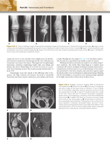
A B C D
Figure 123–5. Various radiologic stages of hemophilic arthropathy. Stages 0 (normal joint) and 1 (fluid in the joint) are not shown. A. Stage 2. Some
osteoporosis and epiphyseal overgrowth are present in knee 2. Epiphysis is wider in knee 2 than in knee 1 (arrows). B. Stage 3. Subchondral bone cysts
(arrowheads). Joint spaces exhibit irregularities. C. Stage 4. Prominent bone cysts with marked narrowing of joint space (arrow). D. Stage 5. Obliteration
of joint space with epiphyseal overgrowth (arrow).
vessels, and nerves. A rare, and often fatal, complication of an abdomi- atrophy. Bleeding into the tongue (Fig. 123–9) or frenulum is particu-
nal hematoma is perforation and drainage into the colon. Subcutaneous larly frequent in young children and usually is caused by trauma.
hematomas may dissect into muscle. Pharyngeal and retropharyngeal Bleeding into fascia and muscle can result in a so-called compart-
hematomas, sometimes complicating simple colds, may enlarge and ment syndrome. This results when hemorrhage in a confined space
obstruct the airway. Hemorrhage in or around the airway is a poten- compresses the arterial vasculature resulting in ischemic muscle injury.
tially life-threatening situation that requires prompt administration of Compartment syndrome tends to occur in the distal part of the extrem-
factor VIII. ities, particularly in the flexor muscles, and sometimes requires urgent
Hemorrhages occur into muscle in the following order of fre- fasciotomy under cover of clotting factor replacement therapy. Bleeding
quency: calf, thigh, buttocks, and forearm. Recurrent or unresolved into the myocardium or erect penis is very unusual, perhaps explained
hematomas may lead to muscle contractures, nerve palsies, and muscle by the high concentration of tissue factor in these tissues.
Figure 123–6. Magnetic resonance imaging (MRI) of normal and
hemophilic knees. A. MRI of normal knee. B. A transverse T2-weighted
spin-echo image of the knee shows an effusion (*) and multiple
foci of hemosiderin deposition (arrows) along the synovium lining
the suprapatellar bursa. C. A sagittal T2-weighted spin-echo image
of the knee shows dark foci of synovial hemosiderin deposition
(white arrows) accompanied by narrowing of the femorotibial joint
(black arrow). D. A sagittal STIR (short tau inversion recovery) image
of the knee (in the same patient as B) demonstrates an effusion in
the suprapatellar bursa (asterisks). The irregular, lumpy surface of the
bursa represents thickened, hemosiderin-laden synovium. Femo-
rotibial joint narrowing (black arrow) is associated with edema in the
subchondral bone of the femoral condyle (white arrow). (Used with
permission of Dr. Jordan Renner, University of North Carolina.)
A B
C D
Kaushansky_chapter 123_p2113-2132.indd 2118 9/21/15 4:36 PM