
Page 327 - Williams Hematology ( PDFDrive )
P. 327
302 Part IV: Molecular and Cellular Hematology Chapter 20: Innate Immunity 303
88
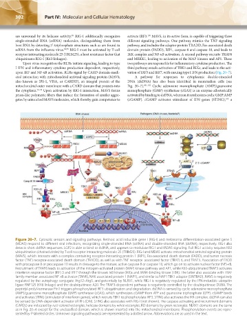
are unwound by its helicase activity. RIG-I additionally recognizes activate IRF3. MAVS, in its active form, is capable of triggering three
100
single-stranded RNA (ssRNA) molecules, distinguishing them from different signaling pathways. One pathway mimics the TNF signaling
host RNA by detecting 5′-triphosphate structures such as are found in pathway, and includes the adaptor protein TRADD, Fas-associated death
ssRNA from the influenza virus. 94,95 RIG-I must be activated by T-cell domain protein (FADD), RIP1, caspase-8 and caspase-10, and leads to
receptor interacting molecule 25 (TRIM25), a host resistance factor that IKK complex and NF-κB activation. A second pathway recruits TRAF6
ubiquitinates RIG-I (K63 linkages). and MEKK1, leading to activation of the MAP kinases and AP1. These
Upon virus recognition the RLHs initiate signaling, leading to type two pathways are responsible for inflammatory cytokine production. The
I IFN and inflammatory cytokine production dependent, respectively, third pathway entails activation of TBK1 and IKKε, and leads to the acti-
upon IRF and NF-κB activation. RLHs signal by CARD domain-medi- vation of IRF3 and IRF7, with ensuing type I IFN production (Fig. 20–7).
ated interaction with mitochondrial antiviral signaling protein (MAVS; A pathway for responses to cytoplasmic double-stranded
also known as IPS-1, VISA, or CARDIF), an integral protein of the DNA (dsDNA) has also been identified in mammalian cells (see
mitochondrial outer membrane with a CARD domain that projects into Fig. 20–7). 101,102 Cyclic adenosine monophosphate (AMP)/guanosine
the cytoplasm. 96–99 Upon activation by RIG-I interaction, MAVS forms monophosphate (GMP) synthetase (cGAS) is an enzyme allosterically
prion-like polymeric fibers that induce the formation of similar aggre- activated by binding to dsDNA, whereon it synthesizes cyclic GMP:AMP
gates by untouched MAVS molecules, which thereby gain competence to (cGAMP). cGAMP activates stimulator of IFN genes (STING), a
103
Figure 20–7. Cytosolic sensors and signaling pathways. Retinoic acid inducible gene I (RIG-I) and melanoma differentiation-associated gene 5
(MDA5) respond to different viral infections, recognizing single-stranded RNA (ssRNA) and double-stranded RNA (dsRNA), respectively. RIG-I also
detects short dsRNA sequences. LGP2 is able to bind to dsRNA, and appears to modulate RIG-I and MDA5 signaling. Full RIG-I activity requires K63
ubiquitination (chained circles) by T-cell receptor interacting molecule 25 (TRIM25). RIG-I and MDA5 activate mitochondrial antiviral signaling protein
(MAVS), which interacts with a complex containing receptor-interacting protein 1 (RIP1), Fas-associated death domain (FADD), and tumor necrosis
factor (TNF) receptor-associated death domain (TRADD), as well as with TNF receptor–associated factor (TRAF) 6, and TRAF3. Association of FADD
with procaspase 8 or procaspase 10 results in cleavage to the mature, active caspase 8 or caspase 10, which go on to activate nuclear factor (NF)-κB.
Recruitment of TRAF6 leads to activation of the mitogen-activated protein (MAP) kinase pathway and AP1, while K63-ubiquitinated TRAF3 activates
interferon response factor (IRF) 3 and IRF7 through the kinases IκB kinase (IKK)ε and TANK-binding kinase (TBK). The latter also associate with TRAF
family member-associated NF-κB activator (TANK), NAK-associated protein 1 (NAP1), and similar to NAP1 TBK1 adaptor (SINTBAD). MAVS is negatively
regulated by the autophagy conjugate Atg12–Atg5, and potentially by NLRX1, while RIG-I is negatively regulated by the IFN-inducible ubiquitin
ligase RNF125 (K48 linkage) and the deubiquitinase A20. The TRAF3-dependent pathway is negatively controlled by the deubiquitinase DUBA. The
peptidyl-prolyl-isomerase Pin1 triggers phosphorylated IRF3 ubiquitination and degradation. dsDNA is sensed by cyclic adenosine monophosphate
(AMP)/guanosine monophosphate (GMP) synthetase (cGAS), which synthesizes cGAMP from ATP and guanosine triphosphate (GTP). cGAMP binds
and activates STING (stimulator of interferon genes), which recruits TBK1 to phosphorylate IRF3. STING also activates the IKK complex. dsDNA can also
be sensed by DNA-dependent activator of IRFs (DAI). STING also associates with RIG-I (not shown). The caspase activating and recruitment domains
(CARDs) are indicated by rust-colored rectangles, helicase domains by white rectangles, RD domains by green rectangles. NLRX1 domains are the same
as in Fig. 20–4 except for the unclassified domain, which is shown inserted into the mitochondrial membrane. Phosphorylation events are repre-
sented by P-labeled circles. Unknown signaling pathway(s) are represented by a dotted arrow. Abbreviations are as used in the text.
Kaushansky_chapter 20_p0293-0306.indd 302 9/17/15 5:52 PM