
Page 498 - Williams Hematology ( PDFDrive )
P. 498
472 Part VI: The Erythrocyte Chapter 31: Structure and Composition of the Erythrocyte 473
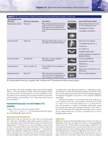
TABLE 31–7. Nomenclature of Red Cell Shapes and Associated Disease States (Continued)
Terminology (Greek
Meaning) Old Terms, Synonyms Description Micrograph Associated Disease States
Drepanocyte (sickle) Sickle cell RBC containing polymerized hemo- Sickle cell disorders (SS, S trait,
globin S; showing varying shapes SC, SD, S thalassemia, etc.)
from bipolar, spiculated forms to Hemoglobin C-Harlem
holly-leaf and irregularly spiculated
forms Hemoglobin Memphis/S
Codocyte (bell) Target cell Bell-shaped RBC that assumes a tar- Obstructive liver disease
get shape on dried films of blood
Hemoglobinopathies (S, C)
Thalassemia
Iron deficiency
Postsplenectomy state
Lecithin cholesterol acetyltrans-
ferase deficiency
Dacryocyte (tear) Teardrop cell RBC with a single elongated or Primary myelofibrosis
pointed extremity
Myelophthisic anemias
Thalassemia
Leptocyte (thin) Thin cell, wafer cell Thin, flat RBC with hemoglobin at Thalassemia
periphery
Obstructive liver disease (± iron
deficiency)
Keratocyte (horn) Horn cell RBC with spicules resulting from DIC or vascular prosthesis
ruptured vacuole; cell appears half-
moon shaped or spindle shaped
DIC, disseminated intravascular coagulation; RBC, red blood cell; TTP, thrombotic thrombocytopenic purpura.
the most aged—they have the highest levels of glycated hemoglobin by fragmentation, with spherocyte formation. A spherogenic mecha-
(HbA ), a very good marker of cell age. The loss of membrane surface nism common to Heinz body hemolytic anemias and immune hemo-
1C
area of the senescent red cells appears to be a result of membrane oxida- lysis is partial phagocytosis of portions of the cell containing aggregates
tion-induced band 3 clustering and consequent membrane vesiculation of denatured hemoglobin and portions of the sensitized membrane,
and the resultant critical decrease in SA:V ratio leads to their removal respectively.
from circulation 59,60 Stomatocytosis appear to be an intermediate form in the gener-
ation of spherocytosis with varying extents of decreased SA:V ratio
PATHOPHYSIOLOGY OF ERYTHROCYTE as a result of loss of membrane surface area or increased cell vol-
SHAPES ume. Stomatocytosis is a feature of hereditary hydrocytosis caused
by increased cell volume and consequent decrease in SA:V ratio. A
Chapter 46 discusses erythrocytes in greater detail. spectrum of abnormal cells varying from normal discocytes to stoma-
See Table 31–7 and Fig. 31–13 for scanning and blood film appear- tocytes, spherostomatocytes, and dense microspherocytes is seen in
ance of pathologically shaped red cells. hereditary spherocytosis.
Spherocytes and Stomatocytes
Spherocytes (Chap. 46) represent red cells, with the most decreased Elliptocytes
SA:V ratio seen in hereditary spherocytosis, immune hemolytic ane- Elliptocytes are seen in hereditary elliptocytosis (Chap. 46) as well as in
mia, stored blood, Heinz body hemolytic anemia, and caused by cell thalassemia (Chap. 48), iron deficiency (Chap. 43), and megaloblastic
fragmentation. 49,61 Stomatocytes are seen in hereditary stomatocytosis, anemia (Chap. 41). In blood films of normal subjects, elliptical or oval
49
as well as in hereditary spherocytosis, alcoholism, cirrhosis, obstructive cells usually constitute less than 1 percent of the erythrocytes. In various
liver disease, and erythrocyte sodium pump defects. 49,62,63 Red cells sen- pathologic situations, with or without anemia (thalassemia trait, folate,
sitized with antibodies, complement, or immune complexes lose cho- and iron deficiency), the number of elliptocytes can increase to 10 per-
lesterol and surface area. As a result, they are less deformable and more cent. Exceptionally, as in dyserythropoiesis, the proportion can be as
osmotically fragile. Heinz body formation leads to membrane depletion high as 50 percent. In hereditary elliptocytosis, the number of elliptical
Kaushansky_chapter 31_p0459-0478.indd 473 9/18/15 10:59 PM