
Page 493 - Williams Hematology ( PDFDrive )
P. 493
468 Part VI: The Erythrocyte Chapter 31: Structure and Composition of the Erythrocyte 469
in the red cell and usually no larger than 0.5 μm in diameter. Howell- cell a characteristic “golf ball–like” appearance when viewed by light
Jolly bodies may be numerous, although generally only one is present. microscopy. Methylene blue and new methylene blue generate a smaller
In pathologic situations, they appear to represent chromosomes that number of variably sized membrane-bound and floating inclusions.
have separated from the mitotic spindle during abnormal mitosis, and These changes are seen most frequently in α-thalassemia but also can
contain a high proportion of centromeric material along with het- be found in patients with unstable hemoglobin (Chap. 49) and in rare
erochromatin. More commonly, during normal maturation they arise patients with primary myelofibrosis who develop acquired hemoglobin
from nuclear fragmentation or incomplete expulsion of the nucleus. H disease.
Howell-Jolly bodies are pitted from the reticulocytes during their tran-
sit through the interendothelial slits of the splenic sinus. They are char- Siderosomes and Pappenheimer Bodies
acteristically present in the blood of splenectomized persons and in Normal or pathologic red cells in blood containing siderosomes (“iron
patients suffering from megaloblastic anemia, and hyposplenic states. bodies”) usually are reticulocytes. The iron granulations are larger and
more numerous in the pathologic state (Chap. 59). Electron microscopy
Pocked (or Pitted) Red Cells shows that many of these bodies are mitochondria containing ferrug-
When viewed by interference-phase microscopy, pocked red cells appear inous micelles rather than the ferritin aggregates characterizing nor-
to have surface membrane “pits” or craters. 37–39 The vesicles or indenta- mal siderocytes. Siderosomes usually are found in the cell periphery,
47
tions characterizing these cells represent autophagic vacuoles adjacent to whereas basophilic stippling tends to be distributed homogeneously
the cell membrane. The vacuoles appear to be instrumental in disposal of throughout the cell. Pappenheimer bodies are siderosomes that stain
cellular debris as the erythrocyte passes through the microcirculation of with Wright stain. Electron microscopy of Pappenheimer bodies shows
the spleen. Within 1 week following splenectomy, pocked red cell counts that the iron often is contained within a lysosome, as confirmed by the
begin to rise, reaching a plateau at 2 to 3 months. Pocked red blood cell presence of acid phosphatase. Siderosomes may contain degenerating
counts sometimes are used as a surrogate test for splenic function. mitochondria, ribosomes, and other cellular remnants.
Cabot Rings STRUCTURE AND SHAPE OF ERYTHROCYTES
The ring-like or figure-of-eight structures sometimes seen in meg-
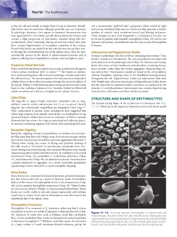
aloblastic anemia within reticulocytes and in an occasional, heavily The normal resting shape of the erythrocyte is a biconcave disc (Fig.
stippled, late-intermediate megaloblast are designated Cabot rings. 40,41 31–12). Variations in the shape and dimensions of the red cell are useful
Their composition is nuclear. Some investigators have suggested that
Cabot rings originate from spindle material that was mishandled during
abnormal mitosis. Others have found no indication of DNA or spindle
filaments but have shown the rings are associated with adherent granu-
lar material containing arginine-rich histone and nonhemoglobin iron.
Basophilic Stippling
Basophilic stippling consists of granulations of variable size and num-
ber that stain deep blue with Wright stain. Electron microscopic studies
42
have shown that punctate basophilia represents aggregated ribosomes.
Clumps form during the course of drying and postvital staining of
the cells, much as “reticulum” in reticulocytes precipitates from ribo-
somes during supravital staining. The clumped ribosomes may include
degenerating mitochondria and siderosomes. In conditions such as lead
intoxication (Chap. 52), pyrimidine 5′-nucleotidase deficiency (Chap.
47), and thalassemia (Chap. 48), the altered reticulocyte ribosomes have
a greater propensity to aggregate. As a result, basophilic granulation
appears larger and is referred to as coarse basophilic stippling.
Heinz Bodies
Heinz bodies are composed of denatured proteins, primarily hemoglo-
bin, that form in red cells as a result of chemical insult; in hereditary
defects of the hexose monophosphate shunt; in the thalassemias (Chap.
43
48); and in unstable hemoglobin syndromes (Chap. 49). Heinz bodies
are not seen on ordinary Wright- or Giemsa-stained blood films. Heinz
bodies are readily visible in red cells stained supravitally with brilliant
cresyl blue or crystal violet and are eliminated as red cells traverse the
endothelial slits of the splenic sinus.
Hemoglobin H Inclusions
Hemoglobin H is composed of β tetramers, indicating that β chains
4
are present in excess as a result of impaired α-chain production (Chap. Figure 31–12. Scanning electron micrographs of distinct red cell
48). Exposure to redox dyes such as brilliant cresyl blue, methylene morphologies. Discoid normal red cells (top left panel). Elliptocytes and
blue, or new methylene blue, results in denaturation and precipitation fragmented red cells (top right panel). Oxygenated sickle red cells (middle
of abnormal hemoglobin. 44–46 Brilliant cresyl blue causes the formation left panel) and deoxygenated sickle red cells (middle right panel). Stoma-
of a large number of small membrane-bound inclusions, giving the tocytic red cells (bottom left panel). Acanthocyte (bottom right panel).
Kaushansky_chapter 31_p0459-0478.indd 468 9/18/15 10:59 PM