
Page 601 - Williams Hematology ( PDFDrive )
P. 601
576 Part VI: The Erythrocyte Chapter 40: Paroxysmal Nocturnal Hemoglobinuria 577
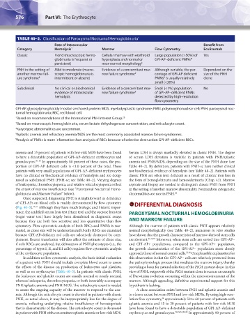
TABLE 40–2. Classification of Paroxysmal Nocturnal Hemoglobinuria *
Rate of Intravascular Benefit from
Category Hemolysis † Marrow Flow Cytometry Eculizumab
Classic Florid (macroscopic hemo- Cellular marrow with erythroid Large population (>50%) of Yes
globinuria is frequent or hyperplasia and normal or GPI-AP–deficient PMNs ¶
persistent) near-normal morphology ‡
PNH in the setting of Mild to moderate (macro- Evidence of a concomitant mar- Although variable, the per- Dependent on the
another marrow fail- scopic hemoglobinuria is row failure syndrome § centage of GPI-AP deficient size of the PNH
ure syndrome § intermittent or absent) PMNs is usually relatively clone
¶
small (<30%)
Subclinical No clinical or biochemical Evidence of a concomitant mar- Small (<1%) population No
evidence of intravascular row failure syndrome § of GPI-AP–deficient PMNs
hemolysis detected by high-resolution
flow cytometry
GPI-AP, glycosylphosphatidylinositol-anchored protein; MDS, myelodysplastic syndrome; PMN, polymorphonuclear cell; PNH, paroxysmal noc-
turnal hemoglobinuria; RBC, red blood cell.
* Based on recommendations of the International PNH Interest Group.
28
† Based on macroscopic hemoglobinuria, serum lactate dehydrogenase concentration, and reticulocyte count.
‡ Karyotypic abnormalities are uncommon.
§ Aplastic anemia and refractory anemia/MDS are the most commonly associated marrow failure syndromes.
¶ Analysis of PMNs is more informative than analysis of RBCs because of selective destruction GPI-AP–deficient RBCs.
anemia and 15 percent of patients with low-risk MDS have been found Serum LDH is always markedly elevated in classic PNH. The degree
to have a detectable population of GPI-AP–deficient erythrocytes and of serum LDH elevation is variable in patients with PNH/aplastic
granulocytes. 30–33 In approximately 80 percent of these cases, the pro- anemia and PNH/MDS, depending on the size of the PNH clone (see
portion of GPI-AP deficient cells is <1.0 percent of the total. These Table 40–2). By definition, patients with PNH-sc have neither clinical
patients with very small populations of GPI-AP–deficient erythrocytes nor biochemical evidence of hemolysis (see Table 40–2). Patients with
have no clinical or biochemical evidence of hemolysis and are desig- classic PNH are often iron deficient as a result of chronic iron loss in
nated as subclinical PNH (PNH-sc; see Table 4 0–2). Varying degrees the form of hemoglobinuria and hemosiderinuria (Chap. 43). Marrow
of leukopenia, thrombocytopenia, and relative reticulocytopenia reflect aspirate and biopsy are needed to distinguish classic PNH from PNH
the extent of marrow insufficiency (see “Paroxysmal Nocturnal Hemo- in the setting of another marrow abnormality. Nonrandom cytogenetic
globinuria and Marrow Failure” below). abnormalities are rare in PNH. 26
Once suspected, diagnosing PNH is straightforward as deficiency
of GPI-APs on blood cells is readily demonstrated by flow cytometry DIFFERENTIAL DIAGNOSIS
(Fig. 40–5). 34,35 Although they have much biologic and historic impor-
tance, the acidified serum lysis test (Ham test) and the sucrose lysis test PAROXYSMAL NOCTURNAL HEMOGLOBINURIA
(sugar water test) have largely been abandoned as diagnostic assays
because they are both less sensitive and less quantitative than flow AND MARROW FAILURE
cytometry. Flow cytometric analysis of both RBCs and PMNs is war- Although the marrow of patients with classic PNH appears relatively
ranted, as clone size will be underestimated if only RBCs are examined normal morphologically (see Table 40–2), numerous in vitro studies
because GPI-AP–deficient red cells are selectively destroyed by com- have shown that the growth characteristics of marrow-derived stem cells
plement. Recent transfusion will also affect the estimate of clone size, are aberrant. 21,36,37 Moreover, when stem cells are sorted into GPI-AP−
if only RBCs are analyzed, but delineation of PNH phenotypes (i.e., the and GPI-AP+ populations, compared to the GPI-AP+ population,
percentage of types I, II, and III cells) requires flow cytometric analysis the growth characteristics of the GPI-AP− population more closely
of the erythrocyte population. approach those of normal control cells. 21,36 One plausible explanation for
In addition to flow cytometric analysis, the basic initial evaluation this observation is that the GPI-AP− cells are relatively protected from
of a patient with PNH should include complete blood count to assess the pathophysiologic process that mediates the marrow injury, thereby
the effects of the disease on production of leukocytes and platelets, providing a basis for natural selection of the PIGA mutant clone. In this
as well as on erythrocytes (Table 40–3). In patients with classic PNH, view of PNH, outgrowth of the PIGA mutant clone is seen as an example
the leukocyte and platelet counts are usually normal or nearly normal, of Darwinian evolution occurring within the microenvironment of the
whereas leukopenia, thrombocytopenia, or both invariably accompany marrow. Although appealing, definitive experimental support for this
PNH/aplastic anemia and PNH/MDS. The reticulocyte count is needed hypothesis is lacking.
to assess the ongoing capacity of the marrow to respond to the ane- A close association exists between PNH and aplastic anemia and
mia. Although the reticulocyte count is elevated in patients with classic to a lesser extent between PNH and low-risk MDSs. By using high-reso-
PNH, as noted above, it may be inappropriately low for the degree of lution flow cytometry, approximately 50 to 60 percent of patients with
34
anemia, reflecting underlying relative insufficiency of hematopoiesis aplastic anemia and 15 to 20 percent of patients with low-risk MDS
that is characteristic of the disease. The reticulocyte count is decreased have been found to have a detectable population of GPI-AP–deficient
in patients with PNH with concomitant aplastic anemia or low-risk MDS. erythrocytes and granulocytes. 30,31,33,38,39 In approximately 90 percent of
Kaushansky_chapter 40_p0571-0582.indd 576 9/17/15 6:22 PM