
Page 600 - Williams Hematology ( PDFDrive )
P. 600
574 Part VI: The Erythrocyte Chapter 40: Paroxysmal Nocturnal Hemoglobinuria 575
TABLE 40–1. Recommendation for Screening Patients for III
Paroxysmal Nocturnal Hemoglobinuria *
History of episodic hemoglobinuria Patient with high percentage of type III
Evidence of nonspherocytic, Coombs-negative intravascular Cell count I cells, high-grade hemolysis
hemolysis (must have high serum lactate dehydrogenase)
Patients with aplastic anemia (screen at diagnosis and once yearly
even in the absence of intravascular hemolysis) A Log fluorescence intensity
Patients with refractory anemia or refractory cytopenias with mul- II
tilineage dysplasia variants of myelodysplastic syndrome †
Patients with venous thrombosis involving unusual sites (usually Patient with high percentage of
have evidence of intravascular hemolysis) Cell count type II cells but low percentage of
• Budd-Chiari syndrome III I type III cells, minimal hemolysis
• Other intraabdominal sites
• Cerebral veins B
• Dermal veins Log fluorescence intensity
I
* Screening by flow cytometric analysis of glycosylphosphatidylinos-
itol-anchored proteins on red blood cells and polymorphonuclear
cells. Cell count Patient with low percentage
of type III cells,minimal
† There is no indication for screening patients with other myelodys- III hemolysis
plastic syndrome classifications.
C Log fluorescence intensity
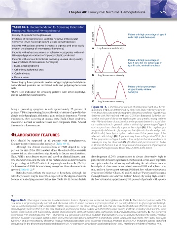
Figure 40–4. Clinical manifestations of paroxysmal nocturnal hemo-
being a presenting symptom in only approximately 25 percent of globinuria (PNH) are determined by clone size and erythrocyte pheno-
patients. Direct questioning frequently elicits a history of episodic dys- type. Mock flow cytometry histograms of erythrocytes from hypothetical
29
phagia and odynophagia, abdominal pain, and male impotence. Venous patients with PNH stained with anti-CD59 are illustrated. Both the pro-
thrombosis, often occurring at unusual sites (Budd-Chiari syndrome, portion and type of abnormal erythrocytes vary greatly among patients
mesenteric, dermal, or cerebral veins), may complicate PNH. Arterial with PNH and these characteristics are important determinants of clini-
thrombosis is less common. cal manifestations. In general, patients with a high percentage of type III
erythrocytes have clinically apparent hemolysis (A). If the erythrocytes
are partially deficient in glycosylphosphatidylinositol-anchored protein
LABORATORY FEATURES (PNH II cells), hemolysis may be modest even if the percentage of the
affected cells is high (B). A patient may have a diagnosis of PNH, but
PNH should be suspected in all patients with nonspherocytic, if the proportion of type III cells is low, only biochemical evidence of
Coombs-negative intravascular hemolysis (Table 40–1). hemolysis may be observed (C). (Modified with permission from Parker
C, Omine M, Richards S, et al: Diagnosis and management of paroxysmal
Although the clinical manifestations of PNH depend in large nocturnal hemoglobinuria. Blood 106(12):3699–3709, 2005.)
part on the size of the PIGA-mutant clone, the extent of the associate
marrow failure also contributes significantly to disease manifestations.
Thus, PNH is not a binary process and based on clinical features, mar- dehydrogenase (LDH) concentration is always abnormally high in
row characteristics, and the size of the mutant clone as determined by patients with clinically significant hemolysis and serves as an important
the percentage of GPI-AP–deficient polymorphonuclear cells (PMNs), surrogate marker for estimating and following the rate of intravascular
the International PNH Interest Group recognizes three disease subcat- hemolysis. A close association exists between PNH and aplastic ane-
egories (Table 40–2). 28 mia, and to a lesser extent between PNH and low-risk myelodysplastic
Reticulocytosis reflects the response to hemolysis, although the syndromes (MDSs) (Chaps. 35 and 87 and see “Paroxysmal Nocturnal
reticulocyte count may be lower than expected for the degree of anemia Hemoglobinuria and Marrow Failure” below). By using high-sensitiv-
because of underlying marrow failure (see Table 40–1). Serum lactate ity flow cytometry, approximately 50 percent of patients with aplastic
Figure 40–3. Phenotypic mosaicism is a characteristic feature of paroxysmal nocturnal hemoglobinuria (PNH). A. The blood of patients with PNH
is a mosaic of phenotypically normal and abnormal cells. In some patients, erythrocytes that are partially deficient in glycosylphosphatidyli-
nositol-anchored proteins (GPI-APs) (called PNH II) are present in the blood along with cells that are completely deficient (PNH III) and cells that are
phenotypically normal (PNH I). In the case illustrated, erythrocytes from a patient with PNH (PNH, upper panels) and from a healthy volunteer (NL, lower
panels) were stained with fluorescently labeled antibodies (anti-CD55, left panels; CD59, right panels) and analyzed by flow cytometry. B. PIGA genotype
determines PNH phenotype. The PNH II phenotype is a consequence of PIGA mutation that partially inactivates enzyme function (red circles), whereas
any PIGA mutation that causes complete loss of enzyme function generates the PNH III phenotype (green, yellow, and blue circles). PNH I cells, have wild-
type PIGA and are the progeny of normal residual hematopoietic stem cells. In a single individual, multiple discrete PIGA mutations can be identified,
accounting for the phenotypic mosaicism based on GPI-AP expression. DAF, decay-accelerating factor; MIRL, membrane inhibitor of reactive lysis.
Kaushansky_chapter 40_p0571-0582.indd 575 9/17/15 6:22 PM