
Page 620 - Williams Hematology ( PDFDrive )
P. 620
594 Part VI: The Erythrocyte Chapter 41: Folate, Cobalamin, and Megaloblastic Anemias 595
A B
C D
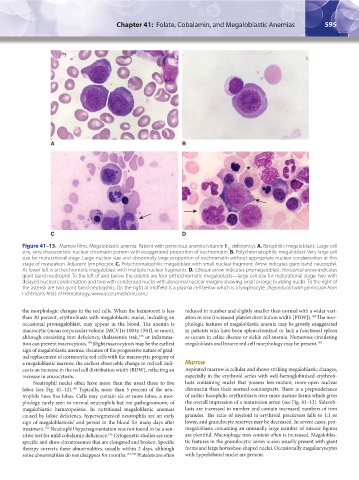
Figure 41–13. Marrow films. Megaloblastic anemia. Patient with pernicious anemia (vitamin B deficiency). A. Basophilic megaloblasts. Large cell
12
size, very characteristic nuclear chromatin pattern with exaggerated proportion of euchromatin. B. Polychromatophilic megaloblast. Very large cell
size for maturational stage. Large nuclear size and abnormally large proportion of euchromatin without appropriate nuclear condensation at this
stage of maturation. Adjacent lymphocyte. C. Polychromatophilic megaloblast with small nuclear fragment. Arrow indicates giant band neutrophil.
At lower left is orthochromatic megaloblast with multiple nuclear fragments. D. Oblique arrow indicates promegaloblast. Horizontal arrow indicates
giant band neutrophil. To the left of and below the asterisk are four orthochromatic megaloblasts—large cell size for maturational stage: two with
delayed nuclear condensation and two with condensed nuclei with abnormal nuclear margins showing small or large budding nuclei. To the right of
the asterisk are two giant band neutrophils. On the right at midfield is a plasma cell below which is a lymphocyte. (Reproduced with permission from
Lichtman’s Atlas of Hematology, www.accessmedicine.com.)
the morphologic changes in the red cells. When the hematocrit is less reduced in number and slightly smaller than normal with a wider vari-
than 20 percent, erythroblasts with megaloblastic nuclei, including an ation in size (increased platelet distribution width [PDW]). The mor-
158
occasional promegaloblast, may appear in the blood. The anemia is phologic features of megaloblastic anemia may be grossly exaggerated
macrocytic (mean corpuscular volume [MCV] is 100 to 150 fL or more), in patients who have been splenectomized or lack a functional spleen
although coexisting iron deficiency, thalassemia trait, or inflamma- as occurs in celiac disease or sickle cell anemia. Numerous circulating
153
tion can prevent macrocytosis. Slight macrocytosis may be the earliest megaloblasts and bizarre red cell morphology may be present. 159
154
sign of megaloblastic anemia. Because of the progressive nature of grad-
ual replacement of normocytic red cells with the macrocytic progeny of
a megaloblastic marrow, the earliest observable change in red cell indi- Marrow
ces is an increase in the red cell distribution width (RDW), reflecting an Aspirated marrow is cellular and shows striking megaloblastic changes,
increase in anisocytosis. especially in the erythroid series with well-hemoglobinized erythrob-
Neutrophil nuclei often have more than the usual three to five lasts containing nuclei that possess less-mature, more-open nuclear
lobes (see Fig. 41–12). Typically, more than 5 percent of the neu- chromatin than their normal counterparts. There is a preponderance
155
trophils have five lobes. Cells may contain six or more lobes, a mor- of earlier basophilic erythroblasts over more mature forms which gives
phology rarely seen in normal neutrophils but not pathognomonic of the overall impression of a maturation arrest (see Fig. 41–13). Siderob-
megaloblastic hematopoiesis. In nutritional megaloblastic anemias lasts are increased in number and contain increased numbers of iron
caused by folate deficiency, hypersegmented neutrophils are an early granules. The ratio of myeloid to erythroid precursors falls to 1:1 or
sign of megaloblastosis and persist in the blood for many days after lower, and granulocyte reserves may be decreased. In severe cases, pro-
5
treatment. Neutrophil hypersegmentation was not found to be a sen- megaloblasts containing an unusually large number of mitotic figures
155
sitive test for mild cobalamin deficiency. Cytogenetic studies are non- are plentiful. Macrophage iron content often is increased. Megaloblas-
156
specific and show chromosomes that are elongated and broken. Specific tic features in the granulocytic series is also usually present with giant
therapy corrects these abnormalities, usually within 2 days, although forms and large horseshoe-shaped nuclei. Occasionally megakaryocytes
some abnormalities do not disappear for months. 151,157 Platelets are often with hyperlobated nuclei are present.
Kaushansky_chapter 41_p0583-0616.indd 595 9/17/15 6:24 PM