
Page 616 - Williams Hematology ( PDFDrive )
P. 616
590 Part VI: The Erythrocyte Chapter 41: Folate, Cobalamin, and Megaloblastic Anemias 591
10
the N ,N -methylene FH reductase reaction. For practical purposes, because cobalamin binds much more tightly to HC than to intrin-
5
4
5
10
90
however, the N ,N -methylene FH reductase reaction is irreversible in sic factor at the acid pH of the stomach. Upon entering the duode-
4
vivo. 81 num, cobalamin is released from the cobalamin–HC protein complex
through digestion by pancreatic proteases, which in normal subjects
FORMATE STARVATION HYPOTHESIS act by selectively degrading HC and the cobalamin–HC complex while
90
This hypothesis holds that formate starvation is the basis for folate- sparing intrinsic factor. Only at this point can cobalamin bind to
intrinsic factor to form the intrinsic factor–cobalamin complex.
responsive megaloblastic anemia of cobalamin deficiency. This theory The intrinsic factor–cobalamin complex, which is very resistant
82
is based on the diminished capacity of cobalamin-deficient lympho- to digestion, traverses the intestine until it reaches the intrinsic factor
91
blasts to incorporate formaldehyde into purine and methionine and receptor, cubilin, a 460-kDa peripheral membrane glycoprotein located
79
92
on experiments showing that N -formyl FH is more effective than FH in the microvillus pits of the ileal mucosa brush-border. Cubilin forms
5
4
4
at correcting some of the abnormalities in folate metabolism seen in part of a multifunctional epithelial receptor complex also found in the
cobalamin deficiency. The hypothesis states that with the decrease in yolk sac and renal proximal tubule cells. In the kidney, it appears to
83
93
methionine production in cobalamin-deficient conditions, the gener- serve a role in the overall body economy through tubular reabsorption
ation of formate is depressed (because normally the methyl group of of cobalamin, but the function of the cubilin receptor complex in the
94
excess methionine is rapidly oxidized to formate), leading to a decline kidney and other polarized epithelial surfaces extends beyond cobala-
84
in the production of N -formyl FH .
5
4 min. The ileal cubilin receptor complex consists of two proteins, cubi-
lin (CUB) and amnionless (AMN), the product of two distinct genes,
INTESTINAL ABSORPTION CUB and AMN. Both proteins, which together have been designated
Intrinsic Factor the “CUBAM complex,” colocalize in the endocytic compartment and
95
Intrinsic factor is one of a number of binding proteins in which cobal- are required for the process of assimilation of cobalamin, AMN serv-
amin is ensconced as it makes its way through the body (Table 41–2). ing as a chaperon for endosomal targeting. Mutations affecting either
Intrinsic factor is needed for the absorption of cobalamins taken orally of the two proteins disrupt the normal process of the intestinal phase
at physiologic dosage levels. Human intrinsic factor is a glycoprotein of cobalamin absorption. In addition to the tightly embracing compo-
85
(Mr approximately 44,000) encoded by a gene on chromosome 11. It nents of the CUBAM complex, a distinct large multifunctional protein,
96
has binding sites for cobalamin and a specific ileal receptor, the former megalin, which belongs to the low-density lipoprotein family, also
situated near the carboxy-terminus and the latter near the aminoter- participates in the conformational changes that accompany internaliza-
minus of the intrinsic factor molecule. Binding to cobalamin is very tion. The concentration of the CUBAM complex rises progressively to
86
97
tight, and involves the 5,6-DMB lower axial ligand of the molecule. This a maximum near the terminal ileum. A specific site on the intrinsic
specificity allows for the exclusion of other noncobalamin corrinoids factor molecule avidly attaches to the receptor in a binding reaction that
2+
64
during the tightly regulated absorptive process. The entrapment of the requires a pH of 5.4 or greater and Ca (or other divalent cations) but
98
vitamin alters the conformation of intrinsic factor, producing a more no energy.
compact form that is resistant to proteolytic digestion. The intrinsic factor–cobalamin receptor complex is taken into
99
In humans, intrinsic factor is synthesized and secreted by the pari- the ileal mucosal cells over 30 to 60 minutes by endocytosis, where
etal cells of the cardiac and fundic mucosa. Secretion of intrinsic fac- the vitamin is processed and released into the portal blood over many
87
tor usually parallels that of hydrochloric acid (HCl). It is enhanced by hours. The receptors recycle to the microvillus surface to shuttle another
99
the presence of food in the stomach, vagal stimulation, histamine, and load of intrinsic factor–cobalamin complex. That this process has a
gastrin. Gastric juice also contains other cobalamin-binding glycopro- limited capacity is evident from estimates of the maximum amount of
88
teins. These proteins were known as the R proteins because of their cobalamin that can be absorbed from a single dose via this physiologic
64,100
rapid electrophoretic mobility compared with intrinsic factor. Elucida- pathway. Defects in the genes that regulate the complex mechanism
tion of the primary protein structure of the R proteins reveals that they of ileal absorption are implicated in autosomal recessive megaloblastic
belong to the same family of isoproteins as the plasma haptocorrin (HC) anemia (MGA1), caused by intestinal malabsorption of cobalamin (see
binder (previously known as transcobalamins I and III). These HC-like “Selective Malabsorption of Cobalamin, Autosomal Recessive Meg-
proteins are produced mainly by the salivary glands. aloblastic Anemia, Imerslund-Gräsbeck Disease” below).
During its sojourn in the ileal enterocyte, the vitamin first appears
Absorption of Cobalamin: Cubilin in the lysosomes, but by 4 hours most of the vitamin is located in the
101
89
Cobalamins in foods are liberated in the stomach by peptic digestion. cytosol. During absorption, the entire intrinsic factor–cobalamin
They are then bound not to intrinsic factor but to the HC-like protein complex appears to be taken into the cell, where the cobalamin is
released while the intrinsic factor is degraded. 102
Cobalamin from a small oral dose (10 to 20 mcg) starts to appear
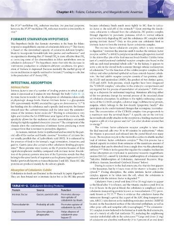
TABLE 41–2. Cobalamin-Binding Proteins in the blood after 3 to 4 hours, and the vitamin reaches a peak level in
6 to 12 hours. In the portal blood, the cobalamin is complexed with a
Protein Source Function cobalamin-transporting protein known as transcobalamin (TC) previ-
103
Intrinsic factor Gastric parietal cells Promotes absorp- ously known as TC II. There is evidence that cobalamin leaves the
tion uptake of enterocyte through a portal that is part of the ABC drug transport sys-
cobalamin by ileum tem, ABCC1 (also known as the multidrug resistance protein1 [MRP1])
Transcobalamin Probably all cells Promotes uptake of located on the basolateral surface of the intestinal epithelium, as well as
104
cobalamin by cells other polar cells and nonpolar cells (macrophages). The cobalamin–
TC complex is now believed to be formed as it exits the ileal enterocyte,
Haptocorrin Exocrine glands, Helps dispose of
phagocytes cobalamin ana- one of a variety of cells that synthesize TC, including the neighboring
105
logues (?) vascular endothelial cells in the submucosa. Large oral doses (1 mg)
of cobalamin are absorbed inefficiently (1 to 2 percent of an oral dose)
Kaushansky_chapter 41_p0583-0616.indd 591 9/17/15 6:24 PM