
Page 621 - Williams Hematology ( PDFDrive )
P. 621
596 Part VI: The Erythrocyte Chapter 41: Folate, Cobalamin, and Megaloblastic Anemias 597
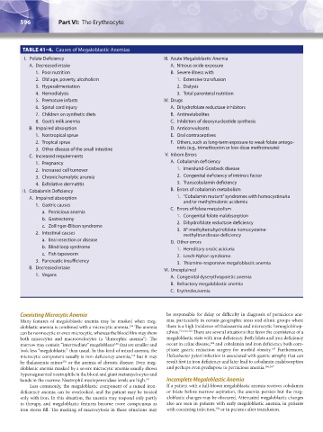
TABLE 41–4. Causes of Megaloblastic Anemias
I. Folate Deficiency III. Acute Megaloblastic Anemia
A. Decreased intake A. Nitrous oxide exposure
1. Poor nutrition B. Severe illness with
2. Old age, poverty, alcoholism 1. Extensive transfusion
3. Hyperalimentation 2. Dialysis
4. Hemodialysis 3. Total parenteral nutrition
5. Premature infants IV. Drugs
6. Spinal cord injury A. Dihydrofolate reductase inhibitors
7. Children on synthetic diets B. Antimetabolites
8. Goat’s milk anemia C. Inhibitors of deoxynucleotide synthesis
B. Impaired absorption D. Anticonvulsants
1. Nontropical sprue E. Oral contraceptives
2. Tropical sprue F. Others, such as long-term exposure to weak folate antago-
3. Other disease of the small intestine nists (e.g., trimethoprim or low-dose methotrexate)
C. Increased requirements V. Inborn Errors
1. Pregnancy A. Cobalamin deficiency
2. Increased cell turnover 1. Imerslund-Gräsbeck disease
3. Chronic hemolytic anemia 2. Congenital deficiency of intrinsic factor
4. Exfoliative dermatitis 3. Transcobalamin deficiency
II. Cobalamin Deficiency B. Errors of cobalamin metabolism
A. Impaired absorption 1. “Cobalamin mutant” syndromes with homocystinuria
1. Gastric causes and/or methylmalonic acidemia
a. Pernicious anemia C. Errors of folate metabolism
b. Gastrectomy 1. Congenital folate malabsorption
c. Zollinger-Ellison syndrome 2. Dihydrofolate reductase deficiency
5
2. Intestinal causes 3. N -methyltetrahydrofolate homocysteine-
methyltransferase deficiency
a. Ileal resection or disease D. Other errors
b. Blind loop syndrome 1. Hereditary orotic aciduria
c. Fish tapeworm 2. Lesch-Nyhan syndrome
3. Pancreatic insufficiency 3. Thiamine-responsive megaloblastic anemia
B. Decreased intake VI. Unexplained
1. Vegans A. Congenital dyserythropoietic anemia
B. Refractory megaloblastic anemia
C. Erythroleukemia
Coexisting Microcytic Anemia be responsible for delay or difficulty in diagnosis of pernicious ane-
Many features of megaloblastic anemia may be masked when meg- mia, particularly in certain geographic areas and ethnic groups where
aloblastic anemia is combined with a microcytic anemia. The anemia there is a high incidence of thalassemia and microcytic hemoglobinop-
154
can be normocytic or even microcytic, whereas the blood film may show athies. 153,162,163 There are several situations that favor the coexistence of a
both microcytes and macroovalocytes (a “dimorphic anemia”). The megaloblastic state with iron deficiency. Both folate and iron deficiency
164
marrow may contain “intermediate” megaloblasts that are smaller and occur in celiac disease, and cobalamin and iron deficiency both com-
160
165
look less “megaloblastic” than usual. In this kind of mixed anemia, the plicate gastric reduction surgery for morbid obesity. Furthermore,
microcytic component usually is iron-deficiency anemia, but it may Helicobacter pylori infection is associated with gastric atrophy that can
154
be thalassemia minor or the anemia of chronic disease. Even meg- result first in iron deficiency and later lead to cobalamin malabsorption
153
aloblastic anemia masked by a severe microcytic anemia usually shows and perhaps even predispose to pernicious anemia. 166,167
hypersegmented neutrophils in the blood and giant metamyelocytes and
bands in the marrow. Neutrophil myeloperoxidase levels are high. 161 Incomplete Megaloblastic Anemia
Less commonly, the megaloblastic component of a mixed iron- If a patient with a full-blown megaloblastic anemia receives cobalamin
deficiency anemia can be overlooked, and the patient may be treated or folate before marrow aspiration, the anemia persists but the meg-
only with iron. In this situation, the anemia may respond only partly aloblastic changes may be obscured. Attenuated megaloblastic changes
to therapy, and megaloblastic features become more conspicuous as also are seen in patients with early megaloblastic anemia, in patients
154
iron stores fill. The masking of macrocytosis in these situations may with coexisting infection, or in patients after transfusion.
Kaushansky_chapter 41_p0583-0616.indd 596 9/17/15 6:24 PM