
Page 619 - Williams Hematology ( PDFDrive )
P. 619
594 Part VI: The Erythrocyte Chapter 41: Folate, Cobalamin, and Megaloblastic Anemias 595
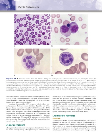
A B
C D
Figure 41–12. A. Pernicious anemia. Blood film. Note the striking oval macrocytes, wide variation in red cell size, and poikilocytes. Despite the
anisocytosis and microcytes, the mean red cell volume is usually elevated, as in this case (mean corpuscular volume [MCV] = 121 fL). B. Marrow pre-
cursors in pernicious anemia. Note very large size of erythroblasts (megaloblasts) and asynchronous maturation. Cell on right is a polychromatophilic
megaloblast with an immature nucleus for that stage of maturation. Cell on left is an orthochromatic megaloblast with a lobulated immature nucleus.
An orthochromatic megaloblast with a condensed nucleus is between and above those two cells. C and D. Two examples of hypersegmented neu-
trophils characteristic of megaloblastic anemia. The morphology of blood and marrow cells in folate-deficient and vitamin B -deficient patients is
12
identical. The extent of the morphologic changes in each case is related to the severity of the vitamin deficiency. (Reproduced with permission from
Lichtman’s Atlas of Hematology, www.accessmedicine.com.)
thymidine fail for the same reason that uridine triphosphate was incor- and intraerythrocytic compensatory changes, it produces few symp-
152
porated into the DNA in the first place. The result is a repetitive itera- toms until the hematocrit is severely depressed. Symptoms, when
tion of flawed DNA repair that ultimately leads to DNA strand breaks, they appear, are those of anemia: weakness, palpitation, fatigue, light-
fragmentation, and apoptotic cell death. 149 headedness, and shortness of breath. The blending of severe pallor and
Addition of deoxyuridine (dU) to marrow cells in culture nor- slight jaundice caused by a combination of intramedullary and extravas-
mally decreases the incorporation of tritiated thymidine into DNA, cular hemolysis produce a characteristic lemon-yellow skin. Leukocyte
because the dU is converted via dUMP→dTMP to unlabeled dTTP, and platelet counts may be low, but rarely cause clinical problems.
which competes with the tritiated thymidine. In megaloblastic cells, this Details of the clinical manifestations are given in the sections on the
effect of added dU is greatly diminished. This finding is consistent with specific forms of megaloblastic anemia later in this chapter.
impairment in the dUMP→dTMP reaction in the megaloblastic cells
and was the basis for the now defunct dU suppression test. The failed LABORATORY FEATURES
150
excision–repair model following dUTP misincorporation into DNA
also explains the chromosome breaks and other abnormalities that Blood Cells
occur in megaloblastic cells. 151 All cell lines are affected. Erythrocytes vary markedly in size and shape,
often are large and oval, and in severe cases can show basophilic stip-
pling and nuclear remnants (Cabot rings and Howell-Jolly bodies). Ery-
CLINICAL FEATURES throid activity in the marrow is enhanced, although the megaloblastic
All megaloblastic anemias share certain general clinical features. Because cells usually die before they are released, accounting for the reduced
the anemia develops slowly, with opportunity for cardiopulmonary reticulocyte count. The more severe the anemia, the more pronounced
Kaushansky_chapter 41_p0583-0616.indd 594 9/17/15 6:24 PM