
Page 618 - Williams Hematology ( PDFDrive )
P. 618
592 Part VI: The Erythrocyte Chapter 41: Folate, Cobalamin, and Megaloblastic Anemias 593
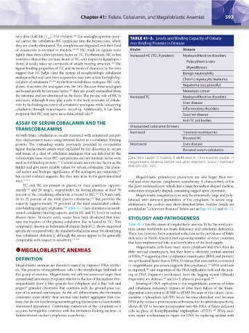
very slow (half-life [T ]: 9 to 10 days). The asialoglycoprotein recep- TABLE 41–3. Levels and Binding Capacity of Cobala-
128
1/2
tor carries the cobalamin–HC complexes into the hepatocytes, where
they are chiefly eliminated. The complexes are degraded, and their load min-Binding Proteins in Disease
of cobalamins is excreted in the bile. 106,129 HC binds its ligands more Binder Disease
tightly than does either intrinsic factor or TC. Furthermore, HC is less Increased HC (TCI, R protein) Myeloproliferative disorders
restrictive than either intrinsic factor or TC with respect to ligand spec-
130
ificity; it avidly takes up corrinoids of widely varying structure. The Polycythemia vera
ligand-binding properties of HC and its mode of clearance by the liver Myelofibrosis
suggest that HC helps clear the system of nonphysiologic cobalamin Benign neutrophilia
analogues that may have been acquired or may have arisen through deg- Chronic myelocytic leukemia
radation of cobalamin. 131,132 As the liver metabolizes analogue–HC com-
plexes, it secretes the analogues into the bile. Because these analogues Hepatoma (occasionally)
130
are bound poorly by intrinsic factor, they are poorly reabsorbed from Metastatic cancer
the intestine and are eliminated in the feces. The precise role of HC is Increased TC Myeloproliferative disorders
unknown, although it may play a role in the body economy of cobala-
min by facilitating excretion of cobalamin analogues while conserving Liver disease
cobalamin through enterohepatic recycling. Additionally, it has been Inflammatory disorders
proposed that HC may serve an antimicrobial role. 64 Gaucher disease
ASSAY OF SERUM COBALAMIN AND THE Anti-TC antibodies
TRANSCOBALAMINS Unsaturated cobalamin binders
As with folate, cobalamin is usually measured with automated compet- Increased Transient neutropenia
itive displacement assays using intrinsic factor as a cobalamin-binding Elevated HC
protein. The misleading results previously provided by competitive Decreased Liver disease
ligand displacement assays were explained by the discovery in serum Elevated serum cobalamin
and tissue of a class of cobalamin analogues that are detected by the
radioisotope assay when HC-type proteins and not intrinsic factor were Data from Lawler S, Roberts P, Hoffbrand A: Chromosome studies in
used as the binding protein. Current assays use intrinsic factor as the megaloblastic anaemia before and after treatment. Scand J Haematol
133
binder and give more reliable values for serum cobalamin. The chem- 8(4):309–320, 1971.
ical nature and biologic significance of the analogues are unknown,
134
but recent evidence suggests that they may arise in the gastrointestinal Megaloblastic granulocyte precursors are also larger than nor-
tract. 131,132 mal and show nuclear-cytoplasmic asynchrony. A characteristic cell is
TC and HC are present in plasma in trace quantities (approx- the giant metamyelocyte, which has a large horseshoe-shaped nucleus,
imately 7 and 20 mcg/L, respectively). In fasting plasma, at least 70 sometimes irregularly shaped, containing ragged open chromatin.
percent of the circulating cobalamin is bound to HC. TC binds only Megaloblastic megakaryocytes may be abnormally large and poly-
135
136
10 to 25 percent of the total plasma cobalamin, but provides the lobated, with deficient granulation of the cytoplasm. In severe meg-
majority (approximately 75 percent) of the total unsaturated cobala- aloblastosis, the nucleus may show detached lobes. Further details are
min-binding capacity of plasma. Table 41–3 lists alterations in unsat- provided in “Laboratory Features” below and in Figs. 41–12 and 41–13.
135
urated cobalamin-binding capacity and in HC and TC levels in various
disease states. In recent years, assays have been developed that mea- ETIOLOGY AND PATHOGENESIS
sure the fraction of the plasma cobalamin that is bound to TC. This
component, known as holotranscobalamin (holoTC), shows improved Table 41–4 lists the causes of megaloblastic anemia. By far the most com-
specificity compared with the standard cobalamin assay for identifying mon causes worldwide are folate deficiency and cobalamin deficiency.
true cobalamin deficiency, although the assays appear to be generally There has, however, been a marked reduction in the prevalence of folate
comparable with respect to sensitivity. 137–143 deficiency in North America and a growing number of other countries
that have implemented folic acid fortification of the food supply.
Megaloblastic cells have much more cytoplasm and RNA than do
MEGALOBLASTIC ANEMIAS their normal counterparts, but they have a relatively normal amount
of DNA, suggesting that cytoplasmic constituents (RNA and protein)
144
DEFINITION are synthesized faster than is DNA. Evidence that maturation is retarded
Megaloblastic anemias are disorders caused by impaired DNA synthe- in megaloblastic precursors supports this conclusion. DNA synthesis
145
sis. The presence of megaloblastic cells is the morphologic hallmark of is impaired, and migration of the DNA replication fork and the join-
146
this group of anemias. Megaloblastic red cell precursors are larger than ing of DNA fragments synthesized from the lagging strand (Okazaki
normal and have more cytoplasm relative to the size of the nucleus. Pro- fragments) are delayed, and the S-phase is prolonged. 146
147
megaloblasts show a blue granule-free cytoplasm and a fine “salt and Slowing of DNA replication in the megaloblastic anemias of folate
pepper” granular chromatin that contrasts with the ground-glass tex- and cobalamin deficiency appears to arise from failure of the folate-
ture of its normal counterpart. As the cell differentiates, the chromatin dependent conversion of dUMP to dTMP. Because of this failure, deox-
condenses more slowly than normal into darker aggregates that coa- yuridine triphosphate (dUTP) levels become abundant and because
lesce, but do not fuse homogeneously, giving the nucleus a characteristic DNA polymerase is promiscuous with respect to its substrate specificity,
fenestrated appearance. Continuing maturation of the cytoplasm as it allows dUTP to become incorporated into the DNA of folate-deficient
acquires hemoglobin contrasts with the immature-looking nucleus—a cells in place of deoxythymidine triphosphate (dTTP). DNA exci-
148
feature termed nuclear-cytoplasmic asynchrony. sion–repair mechanisms to repair the DNA by replacing uridine with
Kaushansky_chapter 41_p0583-0616.indd 593 9/17/15 6:24 PM