
Page 691 - Williams Hematology ( PDFDrive )
P. 691
666 Part VI: The Erythrocyte Chapter 46: Erythrocyte Membrane Disorders 667
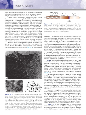
repeats renders the molecule highly flexible and enables it to extend and 24 ANK repeats
condense reversibly, which provides the red cell with elasticity and dura- Band 3, Rh–RhAG- Spectrin- Regulatory
bility to withstand the shear stress encountered in the circulation. binding domain binding domain domain
The core structure of the erythrocyte skeleton consists of spectrin N C
heterotetramers, which are strong but flexible filaments. Tetramers are
assembled from the monomers in a series of events. For initial heterod- Death
imer formation, the α- and β-spectrin chains align in an antiparallel domain
fashion and interact with high affinity through long-range electrostatic Figure 46–5. Schematic of human erythrocyte ankyrin. The N-ter-
33
interactions at a nucleation site, comprising repeats α20–21 and β1–2. minal domain consists of 24 ANK repeats, which bind to band 3 and
This triggers the association of the remaining repeats in the two subun- the Rh–RhAg complex. The central domain attaches to spectrin. The
its in a zipper-like fashion. Repeats at the N-terminus of α-spectrin (αI C-terminal domain varies in different isoforms of ankyrin, which are pro-
domain) and the C-terminus of β-spectrin (βI domain) are the regions duced by alternative splicing of the gene. This domain also contains a
involved in heterodimer self-association to form tetramers. Partial conserved death domain of unknown function.
repeat β17 consists of two helices (A and B) which interact with the
single helix C of partial repeat α0 to form a complete triple helical repeat
(see Fig. 46–3). The interface of this tetramerization site is dominated EF hand of α-spectrin enhances this spectrin-actin-4.1R interaction.
37
34
by hydrophobic contacts supplemented by electrostatic interactions. Numerous other proteins participate in the junctional complex, includ-
Phosphorylation of the C-terminal region of β-spectrin beyond the ing adducin, protein 4.9, p55, tropomodulin and tropomyosin (see Fig.
self-association site decreases the mechanical stability of the membrane. 46–1). Protein 4.1R binds to GPC and band 3, which serves as a sec-
19
At the opposite tail end of the spectrin tetramers, the N terminus of ondary attachment site of the skeleton to integral membrane proteins.
β-spectrin binds to short F-actin filaments, which is potentiated by 4.1R, The main interaction tethering the skeleton to the lipid bilayer is accom-
35
to form the core of a junctional complex, which links six tetramers plished by ankyrin, which links β-spectrin to band 3 (see Fig. 46–1). The
36
together into a hexagonal skeletal network (Fig. 46–4). The C-terminal ankyrin binding site is a flexible pocket formed by repeats 14 and 15 of
β-spectrin near the C-terminal end of the molecule. 38,39 Spectrin also
interacts with phosphatidylserine on the inner leaflet of the lipid bilayer.
Nonrepeat sequences in spectrin provide the recognition sites for
binding to modifiers, including kinases and calmodulin. The functions
of spectrin are to maintain the biconcave disk shape of the red cell, reg-
ulate the lateral mobility of integral membrane proteins, and provide
structural support for the lipid bilayer.
Ankyrin Erythrocyte ankyrin is encoded by the ANK1 gene, which
contains three separate tissue-specific promoters and first exons that are
spliced to a common exon 2. The 206-kDa protein is a versatile bind-
40
ing partner and has three functional domains: an N-terminal 89-kDa
membrane-binding domain, that contains sites for band 3 and other
ligands; a central 62-kDa spectrin-binding domain, and a C-terminal
55-kDa regulatory domain that is responsible for the different iso-
forms of the protein, which influence ankyrin–protein interactions
(Fig. 46–5). 29
The membrane-binding domain contains 24 tandem ankyrin
(ANK) repeats, which are stacked into a superhelical array that is coiled
into a solenoid. This structure behaves like a reversible spring, which
may contribute to the elasticity of the membrane. Each 33-amino-
22
acid ANK repeat is highly conserved and forms an L-shaped struc-
ture composed of two antiparallel α helices separated by a β hairpin.
41
The ANK repeats are connected by unstructured loops and provide an
interface for numerous protein–protein interactions. Erythrocyte ANK
repeats specifically bind to band 3 and the Rh–RhAG macromolecular
complex. 19,29
The spectrin-binding domain contains a small unique subdomain
termed ZU5-ANK, which has a β-strand core with several surface loops
and binds to β-spectrin through hydrophobic and electrostatic inter-
Figure 46–4. Electron micrograph of the human erythrocyte mem- actions. The regulatory domain contains a highly conserved death
42
brane skeleton. Membrane lipids and transmembrane proteins have domain of unknown function in the red cell. The C-terminal section of
been removed and the skeletons were extended during preparation the regulatory domain varies in the different isoforms of ankyrin, pro-
and negative staining to reveal the structure. A. Low-magnification teins 2.1 to 2.6, which are created by alternative splicing and which
43
image reveals an ordered network of proteins. B and C. High-magnifi- exhibit different binding affinities for band 3 and spectrin. Phospho-
cation image and schematic of the hexagonal lattice showing spectrin rylation of ankyrin reduces binding to band 3 and spectrin tetramers.
tetramers (Sp4) and hexamers (Sp6) or double tetramers (2Sp4). Junc-
tional complexes contain actin filaments and protein 4.1R. Globular Protein 4.1R The gene encoding protein 4.1 produces diverse
ankyrin molecules are bound to spectrin tetramers. (Reproduced with isoforms in different tissues and different developmental stages. This
permission from Liu SC et al: Visualisation of the hexagonal lattice in the diversity is accomplished by the use of alternate first exons under the
erythrocyte membrane skeleton. J Cell Biol 104(3):527–536, 1987.) control of different promoters, and alternate initiation codons. This
Kaushansky_chapter 46_p0661-0688.indd 666 9/17/15 6:41 PM