
Page 692 - Williams Hematology ( PDFDrive )
P. 692
666 Part VI: The Erythrocyte Chapter 46: Erythrocyte Membrane Disorders 667
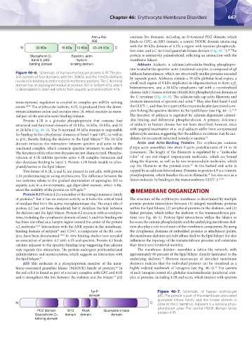
Asn Asp contains five domains, including an N-terminal PDZ domain, which
502 binds to GPC; an SH3 domain; a central HOOK domain interacting
with the 30-kDa domain of 4.1R; a region with tyrosine phosphoryla-
N 30 kDa 16 kDa 10 kDa 22–24 kDa C
48
tion sites; and a C-terminal guanylate kinase domain (Fig. 46–7). The
Glycophorin C, Spectrin, actin protein is extensively palmitoylated, reflecting an interaction with the
band 3, p55- myosin- membrane bilayer.
binding domain binding domain Adducin Adducin, a calcium/calmodulin-binding phosphopro-
tein located at the spectrin–actin junctional complex, is composed of αβ
Figure 46–6. Schematic of human erythrocyte protein 4.1R. The pro- adducin heterodimers, which are structurally similar proteins encoded
tein consists of four domains, with the 30-kDa and the 10-kDa domains by separate genes. Adducins contain a 39-kDa globular head region, a
involved in binding to other red cell membrane proteins. The C-terminal small neck region of 9 kDa implicated in oligomerization to form α β
2 2
domain has an asparagine residue at position 502 in isoform 4.1a, which heterotetramers, and a 30-kDa cytoplasmic tail with a myristoylated
is deamidated in older red cells to form aspartic acid and isoform 4.1b.
alanine-rich C kinase substrate (MARCKS) phosphorylation domain at
the C terminus (Fig. 46–8). The adducin tails cap actin filaments and
49
transcriptional regulation is coupled to complex pre-mRNA splicing promote interaction of spectrin and actin. They also bind band 3 and
events. 44,45 The erythrocyte isoform, 4.1R, is produced from the down- the GLUT-1, and thus form part of the macromolecular junctional com-
stream initiation codon and contains exon 16, which encodes an essen- plex linking the spectrin skeleton to the lipid bilayer (see Fig. 46–1). 21,50
tial part of the spectrin-actin binding domain. The function of adducin is regulated by calcium-dependent calmod-
Protein 4.1R is a globular phosphoprotein that contains four ulin binding and differential phosphorylation. A primary deficiency
structural and functional domains of 30 kDa, 16 kDa, 10 kDa, and 22 of adducin in human disease has not been described; however, mice
to 24 kDa (Fig. 46–6). The N-terminal 30-kDa domain is responsible with targeted inactivation of α- or β-adducin suffer from compensated
for binding to the cytoplasmic domains of band 3 and GPC, as well as spherocytic anemia, suggesting that the adducin mutations may be can-
to p55, thereby linking the skeleton to the lipid bilayer. The 10-kDa didates for recessively inherited hemolytic anemia. 51
19
domain enhances the interaction between spectrin and actin in the Actin and Actin-Binding Proteins The erythrocyte contains
junctional complex, which connects spectrin tetramers to each other. β-type actin assembled into short F-actin protofilaments of 14 to 16
The functions of the other two domains are not characterized. Phospho- monomers. The length of the filaments is regulated by a “molecular
rylation of 4.1R inhibits spectrin–actin–4.1R complex formation and ruler” of two rod-shaped tropomyosin molecules, which are bound
also decreases binding to band 3. Protein 4.1R binds weakly to phos- along the filament, as well as by two tropomodulin molecules, which
52
phatidylserine in the lipid bilayer. cap the filaments at the pointed ends. At the barbed end actin is
Two forms of 4.1R, a and b, are present in red cells, with protein capped by an adducin heterodimer. Dematin or protein 4.9 is a trimeric
53
4.1b predominating in young erythrocytes. The difference between the phosphoprotein, which bundles the actin filaments, but also acts as a
two isoforms relates to the gradual deamidation of asparagine 502 to linker molecule by binding to the transmembrane GLUT-1. 21,50
aspartic acid in a nonenzymatic, age-dependent manner, which influ-
ences the mobility of the protein on SDS gels. 46 MEMBRANE ORGANIZATION
Protein 4.2 Protein 4.2 is a member of the transglutaminase family
of proteins, but it has no enzyme activity as it lacks the critical triad The structure of the erythrocyte membrane is determined by multiple
47
of residues that form the active transglutaminase site. The exact role of protein–protein interactions between (1) integral membrane proteins
protein 4.2 has not been elucidated, but it stabilizes the link between within the lipid bilayer, (2) peripheral proteins in the skeleton, and (3)
the skeleton and the lipid bilayer. Protein 4.2 interacts with several pro- linker proteins, which tether the skeleton to the transmembrane pro-
teins, including the cytoplasmic domain of band 3, and this binding site teins (see Fig. 46–1). Protein–lipid interactions within the bilayer or
has been identified as a hairpin region toward the center of the protein between the anionic phospholipids and the underlying membrane skel-
4.2 molecule. 11,47 Interactions with the ANK repeats in the membrane- eton also play a role in cohesion of the membrane components. By using
47
binding domain of ankyrin and CD47, a component of the Rh com- the cytoplasmic domains of embedded proteins as attachment points,
plex, have been documented. 19,47 In vitro binding studies have revealed the membrane skeleton not only affixes itself to the lipid bilayer but also
an association of protein 4.2 with 4.1R and spectrin. Protein 4.2 binds influences the topology of the transmembrane proteins and constrains
calcium adjacent to the spectrin-binding loop suggesting that calcium their lateral and rotational mobility.
may regulate this interaction. The protein undergoes posttranslational The membrane skeleton resembles a lattice-like network, with
palmitoylation and myristoylation, which suggests an interaction with approximately 60 percent of the lipid bilayer directly laminated to the
36
the lipid bilayer. 47 underlying skeleton. Electron microscopy of stretched membrane
p55 This molecule is a phosphoprotein member of the mem- skeletons indicate that the individual proteins can be visualized as a
36
brane-associated guanylate kinase (MAGUK) family of proteins. In highly ordered meshwork of hexagons (see Fig. 46–4). The corners
48
the red cell it is found as part of a ternary complex with GPC and 4.1R of each hexagon consist of a globular macromolecular junctional com-
and it strengthens the link between the skeleton and the bilayer. p55 plex of proteins, including 4.1R and actin, which interact with spectrin
19
Tyr-P Figure 46–7. Schematic of human erythrocyte
domain p55. The protein is part of the membrane-associated
guanylate kinase family and the kinase domain is
N C close to the C terminus. Adjacent is a tyrosine phos-
phorylation zone. The central HOOK domain binds
PDZ domain SH3 Hook Guanylate kinase protein 4.1R.
Glycophorin C– domain domain domain
binding domain
Kaushansky_chapter 46_p0661-0688.indd 667 9/17/15 6:41 PM