
Page 768 - Williams Hematology ( PDFDrive )
P. 768
742 Part VI: The Erythrocyte Chapter 48: The Thalassemias: Disorders of Globin Synthesis 743
loci on chromosomes 2, 6, and 8, and possibly the X chromosome, at
which polymorphisms are involved in the elevation of fetal hemoglobin
synthesis and that their coinheritance may significantly modify the phe-
notype of different forms of β-thalassemia.
Some mutations that cause β-thalassemia are associated with a
mild phenotype because they result in only modest reduction of β-chain
7
production. For example, mutations at positions –29 and –88 are
+
associated with mild β -thalassemia in Africans. Similarly, particu-
larly mild phenotypes are commonly found with a base substitution at
position 6 in IVS-1 and at position –87 in the 5′-flanking region of the
β-globin gene in Mediterranean populations. The homozygous state
for the IVS-1 position 6 mutation usually produces an extremely mild
form of β-thalassemia. When these “mild” mutations are coinherited
with more-severe β-thalassemia determinants, the compound hete-
rozygous states are characterized by a more severe form of thalassemia
intermedia. Other forms of thalassemia intermedia are associated with
the homozygous state for δβ-thalassemia, the various interactions of β-
thalassemia with δβ-thalassemia, and heterozygous β-thalassemia of the
severe variety or in association with triplicated α-gene loci. 7,10,198 These
complex interactions are the subject of several extensive reviews. 198–200
These mechanisms for the phenotypic variability of the β-thalas-
semias represent only the beginning of our understanding of the genetic
diversity of these conditions. Hence, defining a series of genetic modifi-
192
ers that act at different levels is useful. Primary modifiers represent the
diversity of mutations at the β-globin gene locus. Secondary modifiers
are those, such as α-thalassemia and increased hemoglobin F produc-
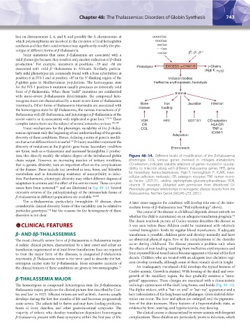
tion, that directly modify the relative degree of the imbalanced globin Figure 48–14. Different levels of modification of the β-thalassemia
chain output. However, an increasing number of tertiary modifiers, phenotype. COL, various genes involved in collagen metabolism;
that is, genetic diversity, have an important effect on the complications CO-selection, indicates variable selection of genes involved in suscep-
of the disease. These include loci involved in iron, bone, and bilirubin tibility to infection along with different thalassemia genes; HFE, gene
metabolism and in determining resistance of susceptibility to infec- for hereditary hemochromatosis; Hgb F, hemoglobin F; ICAM, inter-
tion. Furthermore, phenotypic diversity may reflect different degrees of cellular adhesion molecule; OR, estrogen receptor; TNF, tumor necro-
sis factor; UGT1A1, uridine diphosphate-glucuronyltransferase; VDR,
adaptation to anemia and the effect of the environment. These complex vitamin D receptor. (Adapted with permission from Weatherall DJ:
192
issues have been reviewed and are illustrated in Fig. 48–14. Several Phenotype-genotype relationships in monogenic disease: lessons from the
extensive reviews of the pathophysiology of the intermediate forms of thalassaemias. Nat Rev Genet 2(4):245–255, 2001.)
β-thalassemia in different populations are available. 199,200
The α-thalassemias, particularly hemoglobin H disease, show A later onset suggests the condition will develop into one of the inter-
considerable clinical diversity. Some of this variability can be related to mediate forms of β-thalassemia (see “Pathophysiology” above).
7,41
particular genotypes, but the reasons for the heterogeneity of these The course of the disease in childhood depends almost entirely on
disorders is not clear. whether the child is maintained on an adequate transfusion program.
7,9
The classic textbook picture of Cooley anemia describes the disease as
CLINICAL FEATURES it was seen before these children could be maintained with relatively
normal hemoglobin levels by regular blood transfusions. If adequate
β- AND δβ-THALASSEMIAS transfusion is possible, children grow and develop normally and have
The most clinically severe form of β-thalassemia is thalassemia major. no abnormal physical signs. Few of the complications of the disorder
A milder clinical picture, characterized by a later onset and either no occur during childhood. The disease presents a problem only when
transfusion requirement or at least fewer transfusions than are required the effects of iron loading resulting from ineffective erythropoiesis and
to treat the major form of the illnesses, is designated β-thalassemia from repeated blood transfusions become apparent at the end of the first
intermedia. β-Thalassemia minor is the term used to describe the het- decade. Children who are treated with an adequate iron chelation regi-
erozygous carrier state for β-thalassemia. More extensive accounts of men develop normally, although some of them remain short in height.
the clinical features of these conditions are given in two monographs. 7,9 An inadequately transfused child develops the typical features of
Cooley anemia. Growth is stunted. With bossing of the skull and over-
growth of the maxillary region, the face gradually assumes a “mon-
β-THALASSEMIA MAJOR goloid” appearance. These changes are associated with a characteristic
The homozygous or compound heterozygous state for β-thalassemia, radiologic appearance of the skull, long bones, and hands (Fig. 48–15).
thalassemia major, produces the clinical picture first described by Coo- The diploe widens, with a “hair on end” or “sun ray” appearance and a
ley and Lee in 1925. Affected infants are well at birth. Anemia usually lacy trabeculation of the long bones and phalanges. Gross skeletal defor-
1
develops during the first few months of life and becomes progressively mities can occur. The liver and spleen are enlarged, and the pigmenta-
more severe. The infants fail to thrive and may have feeding problems, tion of the skin increases. Many features of a hypermetabolic state, as
bouts of fever, diarrhea, and other gastrointestinal symptoms. The evidence by fever, wasting, and hyperuricemia, may develop.
majority of infants who develop transfusion-dependent homozygous The clinical course is characterized by severe anemia with frequent
β-thalassemia present with these symptoms within the first year of life. complications. These children are particularly prone to infection, which
Kaushansky_chapter 48_p0725-0758.indd 743 9/18/15 2:58 PM