
Page 103 - Clinical Immunology_ Principles and Practice ( PDFDrive )
P. 103
88 PARt oNE Principles of Immune Response
environmental or other epigenetic events. The HLA-DQB1*06:02 disease, underlying both genetic heritability, but also other
allele on the DRB1*15:01–DQA1*01:02–DQB1*06:02 haplotype factors, such as environmental triggers or epigenetic components.
has been shown to be one of the most important predisposing Multiple genetic loci have been shown to contribute to the risk
genetic factors, with 85–95% of patients with narcolepsy carrying of developing RA. Of these, the HLA class II DRB1 is the most
29
this haplotype. Conversely DQB1*05:01 and DQB1*06:01 have important and contributes 30–50% of the overall genetic sus-
a protective effect. The protective associations of these two DQB1 ceptibility risk.
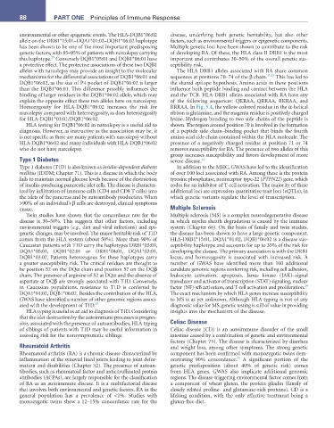
alleles with narcolepsy may provide an insight to the molecular The HLA DRB1 alleles associated with RA share common
mechanisms for the differential associations of DQB1*06:01 and sequences at positions 70–74 of the β chain. 31,32 This has led to
DQB1*06:02, as the size of P4 pocket of DQB1*06:02 is larger the shared epitope hypothesis. Amino acids in these positions
than the DQB1*06:01. This difference possibly influences the influence both peptide binding and contact between the HLA
binding of larger residues in the DQB1*06:02 allele, which may and the TCR. HLA-DRB1 alleles associated with RA have any
explain the opposite effect these two alleles have on narcolepsy. of the following sequences: QKRAA, QRRAA, RKRAA, and
Homozygosity for HLA-DQB1*06:02 increases the risk for RRRAA. In Fig. 5.4, the yellow-colored residue in the α-helical
narcolepsy compared with heterozygosity, as does heterozygosity ribbon is glutamine, and the magenta residue is positively charged
for HLA-DQB1*03:01/DQB1*06:02. lysine. Hydrogen bonding to two side chains of the peptide is
HLA testing for DQB1*06:02 in narcolepsy is a useful aid to shown. The region around position 70 is involved in the formation
diagnosis. However, as instructive as the association may be, it of a peptide side chain–binding pocket that binds the fourth
is not specific as there are many patients with narcolepsy without amino acid side chain contained within the HLA molecule. The
HLA-DQB1*06:02 and many individuals with HLA-DQB1*06:02 presence of a negatively charged residue at position 71 or 74
who do not have narcolepsy. removes susceptibility for RA. The presence of two alleles of this
group increases susceptibility and favors development of more
Type 1 Diabetes severe disease. 33
Type 1 diabetes (T1D) is also known as insulin-dependent diabetes In addition to the MHC, GWAS have led to the identification
mellitus (IDDM; Chapter 71). This is a disease in which the body of over 100 loci associated with RA. Among these is the protein
fails to maintain normal glucose levels because of the destruction tyrosine phosphatase, nonreceptor type-22 (PTPN22) gene, which
of insulin-producing pancreatic islet cells. The disease is character- codes for an inhibitor of T-cell activation. The majority of these
ized by infiltration of immune cells (CD4 and CD8 T cells) into additional loci are expression quantitative trait loci (eQTLs), in
the islets of the pancreas and by autoantibody production. When which genetic variants regulate the level of transcription.
>90% of an individual’s β cells are destroyed, clinical symptoms
ensue. Multiple Sclerosis
Twin studies have shown that the concordance rate for the Multiple sclerosis (MS) is a complex neurodegenerative disease
disease is 30–50%. This suggests that other factors, including in which myelin sheath degradation is caused by the immune
environmental triggers (e.g., diet and viral infections) and epi- system (Chapter 66). On the basis of family and twin studies,
genetic changes, may be involved. The major heritable risk of T1D the disease has been shown to have a large genetic component.
comes from the HLA system (about 50%). More than 90% of HLA-DRB1*15:01, DQA1*01:02, DQB1*06:02 is a disease sus-
Caucasian patients with T1D carry the haplotypes DRB1*03:01, ceptibility haplotype and accounts for up to 35% of the risk for
DQA1*05:01, DQB1*02:01 or DRB1*04:01, DQA1*03:01, developing the disease. The primary association is with the DRB1
DQB1*03:02. Patients heterozygous for these haplotypes carry locus, and homozygosity is associated with increased risk. A
a greater susceptibility risk. The critical residues are thought to number of GWAS have identified more than 100 additional
be position 52 on the DQα chain and position 57 on the DQβ candidate genomic regions conferring risk, including cell adhesion,
chain. The presence of arginine of 52 at DQα and the absence of leukocyte activation, apoptosis, Janus kinase (JAK)–signal
aspartate at DQβ are strongly associated with T1D. Conversely, transducer and activator of transcription (STAT) signaling, nuclear
34
in Caucasian populations, resistance to T1D is conferred by factor (NF)-κB activation, and T-cell activation and proliferation.
DQA1*01:02, DQB1*06:02. Besides the contribution of the HLA, The exact mechanism by which HLA genes increase susceptibility
GWAS have identified a number of other genomic regions associ- to MS is as yet unknown. Although HLA typing is not of any
ated with the development of T1D. 30 diagnostic value for MS, genetic testing is still of value in providing
HLA typing is useful as an aid to diagnosis of T1D. Considering insights into the mechanism of the disease.
that the islet destruction by the autoimmune processes is progres-
sive, associated with the presence of autoantibodies, HLA typing Celiac Disease
of siblings of patients with T1D may be useful information in Celiac disease (CD) is an autoimmune disorder of the small
assessing risk for the nonsymptomatic siblings. intestine caused by a combination of genetic and environmental
factors (Chapter 75). The disease is characterized by diarrhea
Rheumatoid Arthritis and weight loss, among other symptoms. The strong genetic
Rheumatoid arthritis (RA) is a chronic disease characterized by component has been confirmed with monozygotic twins dem-
35
inflammation of the synovial lined joints leading to joint defor- onstrating 90% concordance. A significant portion of the
mation and disabilities (Chapter 52). The presence of autoan- genetic predisposition (about 40% of genetic risk) comes
tibodies, such as rheumatoid factor and anticitrullinated protein from HLA genes. GWAS also implicate additional genomic
antibodies (ACPAs), are largely responsible for the classification regions. The disease-triggering environmental factor comes from
of RA as an autoimmune disease. It is a multifactorial disease a component of wheat gluten, the protein gliadin (family of
that involves both environmental and genetic factors. RA in the closely related proline- and glutamine-rich proteins). CD is a
general population has a prevalence of <1%. Studies with lifelong condition, with the only effective treatment being a
monozygotic twins show a 12–15% concordance rate for the gluten-free diet.