
Page 99 - Clinical Immunology_ Principles and Practice ( PDFDrive )
P. 99
84 PARt oNE Principles of Immune Response
is a positive selection process, whereby only those cells with
TCRs interacting with the self peptide–HLA complex survive.
T cells with receptors that do not recognize any self peptide–HLA
complex are eliminated. The second step is a negative selection
process, whereby among the selected T cells with self recognition,
those with high affinity interactions with the self peptides–
HLA complex are eliminated allowing the rest with lower affinity
interactions to survive and be released in the periphery.
These self peptides constitute the T-cell recognition component
of an individual’s adaptive immune system. This patterning
of TCR recognition on self peptides presented by self MHC
molecules is critical to the development of autoimmunity and
allorecognition.
Evolutionary Considerations Driving the Separate
Functions of Class I and Class II
One basic task of the T cell is to protect the body from two
major types of pathogens: viruses, which would commandeer
the replicative machinery of a cell, and bacteria, which replicate
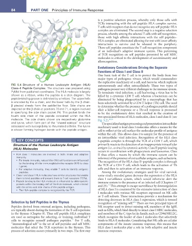
FIG 5.4 Structure of a Human Leukocyte Antigen (HLA) autonomously and often extracellularly. These two types of
Class–II Peptide Complex. The structure was prepared using pathogens present very different challenges to the immune system.
PyMol from published coordinates. The HLA molecule is largely To terminate viral infection, a cell harboring a virus has to be
shown as a ribbon, while the peptide is a stick diagram. The killed by a cytotoxic CD8 T cell, whereas a bacterium can be
peptide-binding groove is delimited by α helices. The upper helix eliminated by being phagocytized by a macrophage that has
is encoded by the α chain, and the lower helix by the β chain. been selectively activated by a CD4 T helper (Th) cell. The need
β pleated sheets form the saddle-like floor. Side chains are to determine whether the presence of a pathogen peptide should
depicted on the β chain at positions 70 and 71, a region involved elicit a killer-cell response or a Th-cell response is presumed
in specifying the side chain pocket P4. This pocket binds the to be the evolutionary drive that resulted in the creation of
fourth side chain of the peptide contained within the HLA two specialized forms of HLA molecules, class I and class II (see
molecule. The side chains shown are respectively glutamine Fig. 5.2). 12,14
and lysine, which form part of the “shared epitope” structure The specialized antigen processing and presentation intracellular
associated with susceptibility to rheumatoid arthritis. The lysine machinery used to load class I molecules offers a means for the
is shown forming hydrogen bonds with the peptide antigen. cell to reflect at the cell surface the molecular profile of antigens
within the cell. This allows class I to sample for the presence of
an intracellular viral infection. Recognition of the HLA class
KEY CoNCEPtS I–peptide complex is through the TCR of a CD8 T cell, which
Structure of the Human Leukocyte Antigen primarily reacts to the detection of an inappropriate intracellular
(HLA) Molecules antigen (i.e., a virus) by cytotoxic activity. Class II peptide loading
occurs in coordination with phagocytosis and lysosomes. Class
• HLA class I molecules are involved in both innate and adaptive II thus offers a means by which the immune system can be
immunity. informed of the presence of extracellular antigens, such as bacteria.
• For innate immunity, natural killer (NK) cell functions are influenced The recognition of the HLA class II–peptide complex is through
by the binding of killer immunoglobulin-like receptor (KIR) to class
I genes. the TCR of a CD4 T cell, which leads to the activation of Th
• For adaptive immunity, they enable T cells to identify antigenic cells and then to activation of an immune response.
peptides. Among the evolutionary strategies used for viral survival,
• Class I and class II HLA molecules have similar structures that enable some virally encoded genes decrease the expression of the HLA
them to bind peptides and present them to T-cell receptors (TCRs). class I surveillance system, which would otherwise alert the
• Peptide binding to the HLA molecule is influenced by allele-specific immune system to the presence of an infected cell (Chapters 17
pockets within the binding cleft of the HLA molecule, which interact 15
with the amino acid side chains of the peptide antigen. and 25). This attempt to escape surveillance by downregulation
• The HLA–peptide complex is recognized by the TCR. of HLA class I is countered by the extensive interaction of class
I molecules with various NK receptors expressed on NK cells
or T-cell subsets. These interactions provide a mechanism for
detecting decreases in HLA class I expression, which is termed
Selection by Self Peptides in the Thymus recognition of “missing self.” There are two principal types of
16
Peptides derived from external antigens, including pathogens, NK receptor used to detect missing self, members of the IgSF,
are typically absent during the formation of the T-cell repertoire such as KIRs, which bind directly to intact class I MHC molecules,
in the thymus (Chapter 8). Thus self peptide–HLA complexes and members of the C-type lectin family, such as CD94/NKG2C,
are used as surrogates for selecting, or training, individual T which recognize the leader of class I molecules that selectively
13
cells to recognize nonself pathogen peptides. For T cells, binds to HLA-E molecules. Considering that NK cells are cellular
“immunological self” is the set of self peptides and self MHC components of the innate immune response, this means that
molecules that select the TCR repertoire in the thymus. This HLA class I molecules play a role in both adaptive and innate
process of selection occurs primarily in two steps. The first step immune responses.