
Page 1031 - Clinical Immunology_ Principles and Practice ( PDFDrive )
P. 1031
994 Part seven Organ-Specific Inflammatory Disease
+
Vitreous humor and from γδ T cells; CD8 Tregs upregulate FasL, IL-10, and
Sclera TGF-β to promote FasL-mediated killing of T cells that recognize
the ocular antigen; TGF-β also induces ocular APCs to secrete
Iris Choroid IL-10 during antigen processing. Apoptotic T cells from the eye
pass in the bloodstream to the spleen, where they are phagocytosed
Lens Retina and induce activation of Th2-type CD4 T cells. The Th2-type
cells control Th1 function by secreting various immunomodula-
tory cytokines. T-cell receptor α-chain fragments from apoptotic
Cornea Vitreous Macula cells are presented in the class I pathway. This event generates
CD8 natural killer (NK) cells, which are capable of deleting the
Anterior Optic CD4 T cells that would otherwise mediate a delayed-type
chamber nerve hypersensitivity response. One study demonstrated that IFN-γ
appears to be important in CD8 Treg suppressive function.
Invariant natural killer T (iNKT) cells, which interact with
Conjunctiva
lipid antigens presented in the context of an MHC-like molecule,
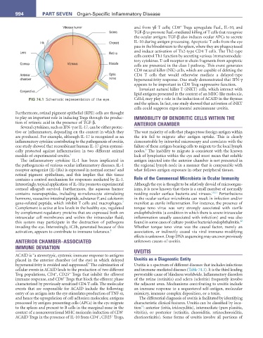
FIG 74.1 Schematic representation of the eye. Cd1d, may play a role in the induction of ACAID in the thymus
and the spleen. In fact, one study showed that activation of iNKT
cells could suppress experimental autoimmune uveitis.
Furthermore, retinal pigment epithelial (RPE) cells are thought
to play an important role in inducing Tregs through the produc- IMMOBILITY OF DENDRITIC CELLS WITHIN THE
tion of retinoic acid in the presence of TGF-β. ANTERIOR CHAMBER
Several cytokines, such as IFN-γ or IL-17, can be either protec-
tive or inflammatory, depending on the context in which they The vast majority of cells that phagocytose foreign antigen within
are produced. For example, although IL-17 is recognized as an the iris fail to migrate after antigen uptake. This is clearly
inflammatory cytokine contributing to the pathogenesis of uveitis, demonstrable by intravital microscopy and correlates with the
one study showed that recombinant human IL-17 given systemi- failure of these antigen-bearing cells to migrate to the local lymph
9
cally protected against inflammation in two different animal nodes. The inability to migrate is consistent with the known
models of experimental uveitis. 5 lack of lymphatics within the eye and must mean that soluble
The inflammatory cytokine IL-1 has been implicated in antigen injected into the anterior chamber is not presented in
the pathogenesis of various ocular inflammatory diseases. IL-1 the regional lymph node in a manner that is comparable with
6
receptor antagonist (IL-1Ra) is expressed in normal cornea and what follows antigen exposure in other peripheral tissues.
retinal pigment epithelium, and this implies that this tissue
contains a control mechanism for responses mediated by IL-1. Role of the Commensal Microbiota in Ocular Immunity
Interestingly, topical application of IL-1Ra promotes experimental Although the eye is thought to be relatively devoid of microorgan-
corneal allograft survival. Furthermore, the aqueous humor isms, it is now known that there is a small number of normally
contains neuropeptides, including α-melanocyte stimulating residing ocular surface bacteria and viruses. 10,11 Perturbations
hormone, vasoactive intestinal peptide, substance P, and calcitonin in the ocular surface microbiota can result in infection and/or
7
gene–related peptide, which inhibit T cells and macrophages. manifest as sterile inflammation. For instance, the presence of
Complement is active at low levels in the healthy eye, regulated torque teno virus was very strongly associated with sterile
by complement regulatory proteins that are expressed both on endophthalmitis (a condition in which there is severe intraocular
intraocular cell membranes and within the intraocular fluid; inflammation usually associated with infection) and was also
12
this system may participate in the destruction of pathogens found in some cases of culture-positive bacterial endophthalmitis.
invading the eye. Interestingly, iC3b, generated because of this Whether torque teno virus was the causal factor, merely an
activation, appears to contribute to immune tolerance. 8 association, or indirectly causal via viral immune-modifying
effects is unknown. Deep DNA sequencing may uncover previously
ANTERIOR CHAMBER–ASSOCIATED unknown causes of uveitis.
IMMUNE DEVIATION
UVEITIS
ACAID is “a stereotypic, systemic immune response to antigens
placed in the anterior chamber (of the eye) in which delayed Uveitis as a Diagnostic Entity
hypersensitivity is avoided and suppressed.” The culmination of Uveitis is a spectrum of different diseases that includes infections
cellular events in ACAID leads to the production of two different and immune-mediated diseases (Table 74.1). It is the third leading
+
+
Treg populations, CD4 , CD25 Tregs that inhibit the afferent preventable cause of blindness worldwide. Inflammatory disorders
+
immune response, and CD8 Tregs that block the efferent phase of the retina (retinitis) and sclera (scleritis) frequently involve
characterized by previously sensitized CD4 T cells. The molecular the adjacent uvea. Mechanisms contributing to uveitis include
events that are responsible for ACAID include the following: an immune response to a sequestered self-antigen, molecular
entry of an antigen into the eye stimulates production of TNF-α, mimicry, immune complex deposition, or a toxin.
and hence the upregulation of cell adhesion molecules; antigens The differential diagnosis of uveitis is facilitated by identifying
processed by antigen-presenting cells (APCs) in the eye migrate characteristic clinical features. Uveitis can be classified by loca-
13
to the spleen and present to B cells in the marginal zone in the tion : anterior (iritis, iridocyclitis), intermediate (pars planitis,
+
context of a nonconventional MHC molecule; induction of CD8 vitritis), or posterior (retinitis, choroiditis, retinochoroiditis,
+
+
ACAID Tregs in the presence of IL-10 from CD4 , CD25 Tregs, chorioretinitis). Some forms of uveitis involve all portions of