
Page 111 - Clinical Immunology_ Principles and Practice ( PDFDrive )
P. 111
CHaPter 6 Overview of T-Cell Recognition 95
β-lactam ring Coupling of a pathogen-derived carbohydrate to another
S CH
CH –CO–NH– CH CH C CH 3 3 foreign protein forms the basis of conjugate vaccines, used to
2
elicit neutralizing antibody responses to bacteria (Chapter 90).
Because related bacterial strains can display distinct polysac-
C N C COOH charide structures that can be recognized by the B cells in the
Penicillin G C H host (serotypes), some conjugate vaccines couple multiple distinct
Nucleophilic attack polysaccharides to a single carrier protein. A classic example is
OH Prevnar® 13, which is composed of polysaccharides from 13
S different serotypes of Streptococcus pneumoniae that are conjugated
Native self protein CH –CO–NH– CH CH C CH 3 to a highly immunogenic inactivated diphtheria toxin protein.
2
CH
+ 3 The conjugate vaccine offers distinct epitopes for the respond-
NH 3
C N C COOH ing B-cell and CD4 T-cell helpers (see Fig. 6.1). The clonally
C O H distributed B-cell Ig receptor binds to the polysaccharides, whereas
the protein carrier provides peptides to be presented on the
Modified surface of the priming dendritic cell (DC) and the antigen-reactive
peptide B cell. This allows focused help by the antigen-specific CD4
Adduct epitope
formation NH T cell.
H O For viral glycoproteins, B cells and CD4 T cells often recognize
COOH C NH C distinct sites on the same protein, with the B cell focusing on
the surface regions of the intact protein structure and the T
CH 3 cell recognizing peptides produced after internalization and
2
CH 3 C S CH CH–CH –CO–NH degradation.
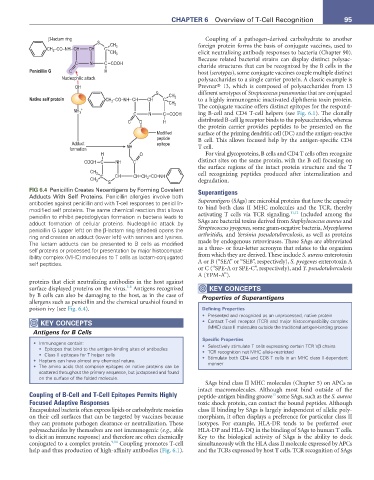
FIG 6.4 Penicillin Creates Neoantigens by Forming Covalent Superantigens
Adducts With Self Proteins. Penicillin allergies involve both
antibodies against penicillin and with T-cell responses to penicillin- Superantigens (SAgs) are microbial proteins that have the capacity
modified self proteins. The same chemical reaction that allows to bind both class II MHC molecules and the TCR, thereby
11,12
penicillin to inhibit peptidoglycan formation in bacteria leads to activating T cells via TCR signaling. Included among the
adduct formation of cellular proteins. Nucleophilic attack by SAgs are bacterial toxins derived from Staphylococcus aureus and
penicillin G (upper left) on the β-lactam ring (shaded) opens the Streptococcus pyogenes, some gram-negative bacteria, Mycoplasma
ring and creates an adduct (lower left) with serines and lysines. arthritidis, and Yersinia pseudotuberculosis, as well as proteins
The lactam adducts can be presented to B cells as modified made by endogenous retroviruses. These SAgs are abbreviated
self proteins or processed for presentation by major histocompat- as a three- or four-letter acronym that relates to the organism
ibility complex (MHC) molecules to T cells as lactam-conjugated from which they are derived. These include S. aureus enterotoxin
self peptides. A or B (“SEA” or “SEB”, respectively), S. pyogenes enterotoxin A
or C (“SPE-A or SPE-C”, respectively), and Y. pseudotuberculosis
A (YPM-A”).
proteins that elicit neutralizing antibodies in the host against
5-8
surface displayed proteins on the virus. Antigens recognized KeY ConCePtS
by B cells can also be damaging to the host, as in the case of Properties of Superantigens
allergens such as penicillin and the chemical urushiol found in
poison ivy (see Fig. 6.4). Defining Properties
• Presented and recognized as an unprocessed, native protein
KeY ConCePtS • Contact T-cell receptor (TCR) and major histocompatibility complex
Antigens for B Cells (MHC) class II molecules outside the traditional antigen-binding groove
Specific Properties
• Immunogens contain: • Selectively stimulate T cells expressing certain TCR Vβ chains
• Epitopes that bind to the antigen-binding sites of antibodies
• Class II epitopes for T helper cells • TCR recognition not MHC allele-restricted
• Haptens can have almost any chemical nature. • Stimulate both CD4 and CD8 T cells in an MHC class II-dependent
• The amino acids that comprise epitopes on native proteins can be manner
scattered throughout the primary sequence, but juxtaposed and found
on the surface of the folded molecule.
SAgs bind class II MHC molecules (Chapter 5) on APCs as
intact macromolecules. Although most bind outside of the
Coupling of B-Cell and T-Cell Epitopes Permits Highly peptide-antigen binding groove some SAgs, such as the S. aureus
13
Focused Adaptive Responses toxic shock protein, can contact the bound peptides. Although
Encapsulated bacteria often express lipids or carbohydrate moieties class II binding by SAgs is largely independent of allelic poly-
on their cell surfaces that can be targeted by vaccines because morphism, it often displays a preference for particular class II
they can promote pathogen clearance or neutralization. These isotypes. For example, HLA-DR tends to be preferred over
polysaccharides by themselves are not immunogenic (e.g., able HLA-DP and HLA-DQ in the binding of SAgs to human T cells.
to elicit an immune response) and therefore are often chemically Key to the biological activity of SAgs is the ability to dock
conjugated to a complex protein. 9,10 Coupling promotes T-cell simultaneously with the HLA class II molecule expressed by APCs
help and thus production of high-affinity antibodies (Fig. 6.1). and the TCRs expressed by host T cells. TCR recognition of SAgs