
Page 128 - Clinical Immunology_ Principles and Practice ( PDFDrive )
P. 128
CHaPTEr 7 B-Cell Development and Differentiation 111
is also known as heat-stable antigen (HSA). Mice made deficient PIP-deficient mice lack GCs in peripheral lymphoid organs and
in CD24 show a leaky block in B-cell development with a reduc- exhibit defects in B-cell activation.
tion in late preB-cell and immature B-cell populations. However, Ikaros and Aiolos belong to the same zinc finger transcription
peripheral B-cell numbers are normal, and no impairment of factor family. Although both are expressed during lymphoid
immune function has been demonstrated. development, Ikaros is expressed in stem cells and mature
CD38 is a bifunctional enzyme that can synthesize cyclic lymphocytes, whereas Aiolos is only expressed after commit-
adenosine diphosphate–ribose (cADPR) from nicotinamide ment to the B-cell lineage. Ikaros transcripts are subject to
+
adenine dinucleotide (NAD ) and also hydrolyze cADPR to alternate splicing. It can generate several isoforms, each of which
ADP-ribose. It is presumed that the enzyme exists to ADP- differs in its DNA-binding pattern, tendency to dimerize, and
ribosylate target molecules. CD38 is expressed on preB cells, nuclear localization. Among the genes bound by Ikaros are
activated B cells, and early plasma cells, but not on immature TdT, λ14.1 (λ5), VpreB, and LCK. Ikaros-deficient mice lack
or mature B cells or on mature plasma cells. Antibodies to CD38 B cells. Aiolos-deficient mice exhibit elevated levels of IgG and
can inhibit B lymphopoiesis, induce B-cell proliferation, and IgE. As they age, these mice tend to develop autoantibodies and
protect B cells from apoptosis. CD38 knock-out mice exhibit B-cell lymphomas.
marked deficiencies in antibody responses to T cell–dependent The E2A locus encodes two basic helix–loop–helix transcrip-
protein antigens and augmented antibody responses to T-cell– tion factors that represent two alternately spliced products, E12
independent type 2 polysaccharide antigens. and E47. Targets for E2A include RAG-1 and TdT. Although the
functions of E12 and E47 overlap, E47 appears to play the greater
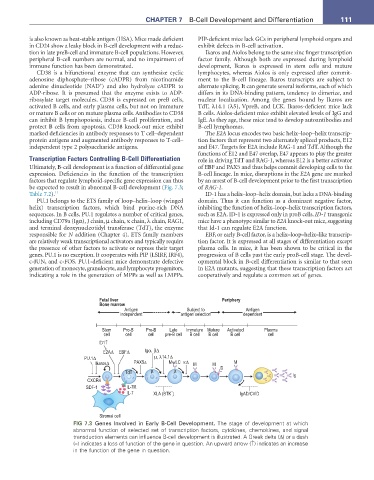
Transcription Factors Controlling B-Cell Differentiation role in driving TdT and RAG-1, whereas E12 is a better activator
Ultimately, B-cell development is a function of differential gene of EBF and PAX5 and thus helps commit developing cells to the
expression. Deficiencies in the function of the transcription B-cell lineage. In mice, disruptions in the E2A gene are marked
factors that regulate lymphoid-specific gene expression can thus by an arrest of B-cell development prior to the first transcription
be expected to result in abnormal B-cell development (Fig. 7.3; of RAG-1.
Table 7.2). 11 ID-1 has a helix–loop–helix domain, but lacks a DNA-binding
PU.1 belongs to the ETS family of loop–helix–loop (winged domain. Thus it can function as a dominant negative factor,
helix) transcription factors, which bind purine-rich DNA inhibiting the function of helix–loop–helix transcription factors,
sequences. In B cells, PU.1 regulates a number of critical genes, such as E2A. ID-1 is expressed only in proB cells. ID-1 transgenic
including CD79a (Igα), J chain, µ chain, κ chain, λ chain, RAG1, mice have a phenotype similar to E2A knock-out mice, suggesting
and terminal deoxynucleotidyl transferase (TdT), the enzyme that Id-1 can regulate E2A function.
responsible for N addition (Chapter 4). ETS family members EBF, or early B-cell factor, is a helix–loop–helix–like transcrip-
are relatively weak transcriptional activators and typically require tion factor. It is expressed at all stages of differentiation except
the presence of other factors to activate or repress their target plasma cells. In mice, it has been shown to be critical in the
genes. PU.1 is no exception. It cooperates with PIP (LSIRF, IRF4), progression of B cells past the early proB-cell stage. The devel-
c-JUN, and c-FOS. PU.1-deficient mice demonstrate defective opmental block in B-cell differentiation is similar to that seen
generation of monocyte, granulocyte, and lymphocyte progenitors, in E2A mutants, suggesting that these transcription factors act
indicating a role in the generation of MPPs as well as LMPPs. cooperatively and regulate a common set of genes.
Fetal liver Periphery
Bone marrow
Antigen Subject to Antigen
independent antigen selection dependent
Stem Pro-B Pre-B Late Immature Mature Activated Plasma
cell cell cell pre-B cell B cell B cell B cell cell
ID1↑
E2A∆ EBF∆ Igα, β∆
PU.1∆ µ, λ14.1∆
Ikaros∆ PAX5∆ MψLC κ∆ M M D M
TdT µ µ
Ig
CXCR4
SDF-1 IL-7R
IL-7 XLA (BTK ) - IgAD/CVID
Stromal cell
FIG 7.3 Genes Involved in Early B-Cell Development. The stage of development at which
abnormal function of selected set of transcription factors, cytokines, chemokines, and signal
transduction elements can influence B-cell development is illustrated. A Greek delta (Δ) or a dash
(–) indicates a loss of function of the gene in question. An upward arrow (↑) indicates an increase
in the function of the gene in question.