
Page 131 - Clinical Immunology_ Principles and Practice ( PDFDrive )
P. 131
114 ParT ONE Principles of Immune Response
B-cell follicle
Mantle zone T cell–dependent activation of B cells
Germinal center formation
Network
of FDC
Light zone – affinity selected differentiation into Memory cells
Dark zone – B-cell proliferation and diversification Plasma cells
T-cell zone
Ti antigen activation of marginal zone B cells
Extrafollicular differentiation into short-lived plasma cells
Marginal zone Macrophages bring the antigen to the marginal zone
Dendritic cells bring antigen to the T-cell zone
Marginal zone B cells bring antigen to the follicle
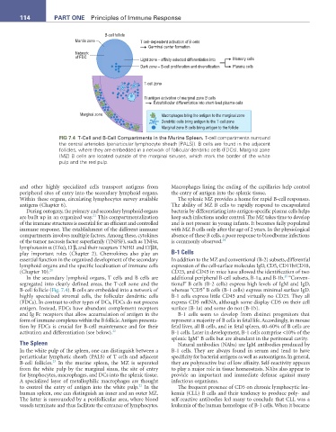
FIG 7.4 T-Cell and B-Cell Compartments in the Murine Spleen. T-cell compartments surround
the central arterioles (periarticular lymphocyte sheath [PALS]). B cells are found in the adjacent
follicles, where they are embedded in a network of follicular dendritic cells (FDCs). Marginal zone
(MZ) B cells are located outside of the marginal sinuses, which mark the border of the white
pulp and the red pulp.
and other highly specialized cells transport antigens from Macrophages lining the ending of the capillaries help control
peripheral sites of entry into the secondary lymphoid organs. the entry of antigen into the splenic tissue.
Within these organs, circulating lymphocytes survey available The splenic MZ provides a home for rapid B-cell responses.
antigens (Chapter 6). The ability of MZ B cells to rapidly respond to encapsulated
During ontogeny, the primary and secondary lymphoid organs bacteria by differentiating into antigen-specific plasma cells helps
21
are built up in an organized way. This compartmentalization keep such infections under control. The MZ takes time to develop
of the immune structures is essential for an efficient and controlled and is not present in young infants. It becomes fully populated
immune response. The establishment of the different immune with MZ B cells only after the age of 2 years. In the physiological
compartments involves multiple factors. Among these, cytokines absence of these B cells, a poor response to bloodborne infections
of the tumor necrosis factor superfamily (TNFSF), such as TNFα, is commonly observed. 24
lymphotoxin α (LTα), LTβ, and their receptors TNFR1 and LTβR,
play important roles (Chapter 2). Chemokines also play an B-1 Cells
essential function in the organized development of the secondary In addition to the MZ and conventional (B-2) subsets, differential
lymphoid organs and the specific localization of immune cells expression of the cell-surface molecules IgD, CD5, CD11b/CD18,
(Chapter 10). 20 CD23, and CD45 in mice have allowed the identification of two
25
In the secondary lymphoid organs, T cells and B cells are additional peripheral B-cell subsets, B-1a, and B-1b. “Conven-
segregated into clearly defined areas, the T-cell zone and the tional” B cells (B-2 cells) express high levels of IgM and IgD,
B-cell follicle (Fig. 7.4). B cells are embedded into a network of whereas “CD5” B cells (B-1 cells) express minimal surface IgD.
highly specialized stromal cells, the follicular dendritic cells B-1 cells express little CD45 and virtually no CD23. They all
(FDCs). In contrast to other types of DCs, FDCs do not process express CD5 mRNA, although some display CD5 on their cell
antigen. Instead, FDCs have abundant complement receptors surface (B-1a) and some do not (B-1b).
and Ig Fc receptors that allow accumulation of antigen in the B-1 cells seem to develop from distinct progenitors that
form of immune complexes within the B follicle. Antigen presenta- represent a majority of B cells in fetal life. Accordingly, in mouse
tion by FDCs is crucial for B-cell maintenance and for their fetal liver, all B cells, and in fetal spleen, 40–60% of B cells are
activation and differentiation (see below). 22 B-1 cells. Later in development, B-1 cells comprise <10% of the
+
splenic IgM B cells but are abundant in the peritoneal cavity.
The Spleen Natural antibodies (NAbs) are IgM antibodies produced by
In the white pulp of the spleen, one can distinguish between a B-1 cells. They are always found in serum and tend to have
periarticular lymphatic sheath (PALS) of T cells and adjacent specificity for bacterial antigens as well as autoantigens. In general,
23
B-cell follicles. In the murine spleen, the MZ is separated they are polyreactive but of low affinity. Self-reactivity appears
from the white pulp by the marginal sinus, the site of entry to play a major role in tissue homeostasis. NAbs also appear to
for lymphocytes, macrophages, and DCs into the splenic tissue. provide an important and immediate defense against many
A specialized layer of metallophilic macrophages are thought infectious organisms.
22
to control the entry of antigen into the white pulp. In the The frequent presence of CD5 on chronic lymphocytic leu-
human spleen, one can distinguish an inner and an outer MZ. kemia (CLL) B cells and their tendency to produce poly- and
The latter is surrounded by a perifollicular area, where blood self-reactive antibodies led many to conclude that CLL was a
vessels terminate and thus facilitate the entrance of lymphocytes. leukemia of the human homologue of B-1 cells. When it became