
Page 1284 - Clinical Immunology_ Principles and Practice ( PDFDrive )
P. 1284
1246 Part ElEvEn Diagnostic Immunology
Flow cytometry can be used to test for the presence or absence CD62L, and CXCR7 and memory T cells that express the alterna-
of a specific cell surface antigen. An example of this type of tive CD45 isoform, CD45RO (and varied CD62L, or CXCR7,
application is in the evaluation of a patient with a history of depending on whether the cells are central or effector memory
35
recurrent skin infections, delayed wound healing, and persistent cells). In addition, memory B cells can be detected by the
granulocytosis, which suggests a diagnosis of leukocyte adhesion expression of CD27 and can be further divided into isotype-
deficiency type 1 (LAD-1). 28,32 This disorder results from a defect switched and isotype-nonswitched memory cells on the basis
in the gene encoding CD18, preventing the expression of three of their pattern of surface immunoglobulin expression. 29,36
different heterodimeric adhesion molecules (β 2 integrins) each Defects associated with familial lymphohistiocytosis (FLH)
containing CD18 (Chapter 22). This disorder can usually be are generally associated with abnormal NK cell function. Many
diagnosed by studying granulocytes (and lymphocytes) for the of the FHL-causing defects can be determined by flow cytometry.
expression of CD18 (as well as the three isoforms of CD11). For example, signaling lymphocyte activation molecule (SLAM)
Patients often have decreased rather than absent CD18 expression associated protein (SAP) and X-linked inhibitor of apoptosis
and confirmation of the diagnosis can be accomplished by (XIAP) intracellular staining can be used to evaluate for X-linked
demonstrating failure of CD18 (and CD11a, 11b, 11c) upregula- lymphoproliferative (XLP) disorders types 1 and 2, respectively.
tion following granulocyte activation. 28 Likewise, lack of intracellular perforin expression in NK cells
Immunophenotyping can also help address questions regarding would be indicative of hemophagocytic lymphohistiocytosis
the level of cell differentiation. Antibodies specific to proteins (HLH) type 2. Additionally, the evaluation of CD107a surface
expressed by early (precursor) cells represent one approach and expression, which is normally expressed on cytoplasmic granules
would include evaluating for the thymocyte marker CD1 or the and upon incubation with specific target cells (e.g., K562 cells)
pre-B-cell marker CD10 (CALLA), to cite a few. However, defining gets expressed on the surface of NK cells, is useful in determining
the developmental level of a particular cell population or sub- the underlying genetic defect causing FHL. 37,38 Specifically, lack
population is best accomplished by using a panel of reagents of CD107a upregulation is suggestive of syntaxin-11 or
that span the natural history of the cell lineage. This approach MUNC-13.4 defects.
represents the standard for testing leukemias and lymphomas.
In addition to defining the presence or absence of specific antigens, INTRACELLULAR EVALUATION
evaluating their level of expression is also valuable, which may
be altered in abnormal cells. Malignant cells may also express Cellular Activation
antigens associated with different lineages and have altered FSC
and SSC characteristics, as well as diminished or absent CD45 ClInICal rElEvanCE
expression, requiring modified gating approaches.
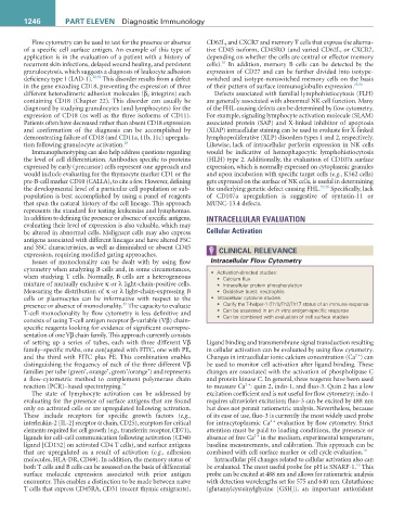
Issues of monoclonality can be dealt with by using flow Intracellular Flow Cytometry
cytometry when analyzing B cells and, in some circumstances, • Activation-directed studies:
when studying T cells. Normally, B cells are a heterogeneous • Calcium flux
mixture of mutually exclusive κ or λ light-chain-positive cells. • Intracellular protein phosphorylation
Measuring the distribution of κ or λ light-chain-expressing B • Oxidative burst: neutrophils
cells or plasmocytes can be informative with respect to the • Intracellular cytokine studies:
33
presence or absence of monoclonality. The capacity to evaluate • Clarify the T-helper-1 (Th1)/Th2/Th17 status of an immune response
T-cell monoclonality by flow cytometry is less definitive and • Can be assessed in an in vitro antigen-specific response
consists of using T-cell antigen receptor β-variable (Vβ) chain- • Can be combined with evaluation of cell surface studies
specific reagents looking for evidence of significant overrepre-
sentation of one Vβ chain family. This approach currently consists
of setting up a series of tubes, each with three different Vβ Ligand binding and transmembrane signal transduction resulting
family–specific mAbs, one conjugated with FITC, one with PE, in cellular activation can be evaluated by using flow cytometry.
2 +
and the third with FITC plus PE. This combination enables Changes in intracellular ionic calcium concentration (Ca ) can
distinguishing the frequency of each of the three different Vβ be used to monitor cell activation after ligand binding. These
+
+
+
+
families per tube (green , orange , green /orange ) and represents changes are associated with the activation of phospholipase C
a flow-cytometric method to complement polymerase chain and protein kinase C. In general, three reagents have been used
2 +
reaction (PCR)–based spectratyping. 34 to measure Ca : quin 2, indo-1, and fluo-3. Quin 2 has a low
The state of lymphocyte activation can be addressed by excitation coefficient and is not useful for flow cytometry; indo-1
evaluating for the presence of surface antigens that are found requires ultraviolet excitation; fluo-3 can be excited by 488 nm
only on activated cells or are upregulated following activation. but does not permit ratiometric analysis. Nevertheless, because
These include receptors for specific growth factors (e.g., of its ease of use, fluo-3 is currently the most widely used probe
2 +
interleukin-2 [IL-2] receptor α chain, CD25), receptors for critical for intracytoplasmic Ca evaluation by flow cytometry. Strict
elements required for cell growth (e.g., transferrin receptor, CD71), attention must be paid to loading conditions, the presence or
2 +
ligands for cell–cell communication following activation (CD40 absence of free Ca in the medium, experimental temperature,
ligand [CD152] on activated CD4 T cells), and surface antigens baseline measurements, and calibration. This approach can be
that are upregulated as a result of activation (e.g., adhesion combined with cell surface marker or cell cycle evaluation. 10
molecules, HLA-DR, CD69). In addition, the memory status of Intracellular pH changes related to cellular activation also can
10
both T cells and B cells can be assessed on the basis of differential be evaluated. The most useful probe for pH is SNARF-1. This
surface molecule expression associated with prior antigen probe can be excited at 488 nm and allows for ratiometric analysis
encounter. This enables a distinction to be made between naïve with detection wavelengths set for 575 and 640 nm. Glutathione
T cells that express CD45RA, CD31 (recent thymic emigrants), (glutamylcysteinylglycine [GSH]), an important antioxidant