
Page 1339 - Clinical Immunology_ Principles and Practice ( PDFDrive )
P. 1339
1300 ParT ElEvEN Diagnostic Immunology
DESCRIPTION AND PRINCIPLES OF DNA Fixed cells
DIAGNOSTIC TECHNIQUES
The overriding theme of all DNA diagnostic techniques is the
detection of variants in the DNA molecule that are associated
with disease. This has historically involved a targeted search in Denature DNA in situ
one or a few specific genes or regions, but the field is evolving
rapidly toward techniques that allow for simultaneous survey
of the entire genome. DNA copy number analysis and DNA
sequencing have come to dominate individual genetic diagnostic
methods. Bioinformatics is a necessary diagnostic laboratory
discipline that must be coupled to these methods, particularly Hybridize
when the whole genome is surveyed.
DIG-dUTP labeled
Detection of Disease-Causing Copy Number Variants: DNA probe
FISH and Microarrays
Many genetic diseases are caused by deletions or duplications
in human DNA. These conditions typically have copy number
abnormalities ranging from 500 kilobases (Kb) to 5 megabases Fluorescent-tagged
(Mb) and are sometimes referred to as “genomic disorders.” The anti-DIG antibody
most common genomic disorder with a clinically important
effect on the immune system is 22q11 deletion syndrome. This
disorder affects about 1 per 4000 live-born infants and accounts
for about 90% of DGS cases. The karyotype, in which a metaphase
chromosome spread was stained and visualized under the Fluorescence Microscopy
microscope, was for many decades the only clinically useful test
for the detection of copy number changes. The karyotype offered
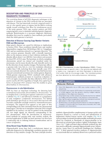
a genome-wide view, but at a very low resolution: alterations FIG 96.2 Fluorescence in situ Hybridization (FISH). Probes
smaller than 5 Mb, including those that typically cause DGS, complementary to specific sites in the genome are labeled with
were not routinely detected. Two methods, described below, have a hapten (e.g., digoxigenin) and then hybridized to denatured
replaced conventional karyotypes for routine genetic diagnosis. DNA within cells on microscope slides. The hybridized probes
Whole-genome sequencing (WGS) combined with advanced are then detected by immunofluorescence microscopy.
bioinformatics methods promises to transform evaluation of
CNVs yet again. In principle, the data collected in WGS can be
used to resolve all CNVs regardless of length, thus capturing all KEY CONCEPTS
forms of structural DNA variation from the smallest to gross
abnormalities in chromosomes. Molecular Methods
Fluorescence in situ Hybridization • Most DNA diagnostics rely on DNA copy number analysis or DNA
sequencing.
FISH has been an important technology for detecting both • Polymerase chain reaction (PCR) allows for exponential amplification
19
constitutional and somatic abnormalities in chromosomes. In of a DNA segment from the genome, allowing multiple analytical
its simplest form, a cloned gene segment (referred to as a probe) procedures on the single pure sequence.
is labeled (Fig. 96.2) and allowed to hybridize to a preparation • Array-based copy number analysis has become the first-line method
for finding small abnormalities in the structure of chromosomes.
of early metaphase (condensed) chromosomes. The labeled • New high-throughput sequencing methods have emerged as practical
probe hybridizes specifically to a locus and is then detected with alternatives to older single analysis approaches.
secondary antibodies and high-sensitivity immunofluorescence • Bioinformatics is an emerging discipline relevant to molecular diagnosis.
techniques. The targeted locus appears as two pairs of dots Integration of bioinformatics techniques plays a necessary role in data
corresponding to the sister chromatids from the two chromosomal acquisition and interpretation.
homologues. Cells that are in interphase (when the DNA is largely
unfolded in the nucleus) can also be probed. In the interphase
cell, two single signals are observed corresponding to the synthetic oligonucleotides complementary to human nucleic acid
two chromosomes. With the introduction of microarray- sequences bound to a surface or bead, and hybridization of
based methods for genome-wide copy number analysis, the fragmented, labeled human nucleic acid samples (Fig. 96.3).
practical application of FISH has been narrowed but does have Array printing of activated phosphoramidite nucleotide precursors
a continuing role in defining balanced translocations and in allows for hundreds to thousands of oligonucleotides to be
cancer cytogenetics. fabricated on activated glass surfaces (Agilent Technologies, Santa
20
Clara, CA). Light-activated oligonucleotide synthesis has also
Array-Based Copy Number Analysis been used to efficiently customize oligonucleotide arrays (Affyme-
Oligonucleotide arrays have had a revolutionary impact on trix Inc., Santa Clara, CA; and Roche Nimblegen Inc., Madison,
21
molecular diagnostics, by offering high-resolution, genome-wide WI). A third approach involves synthesis of oligonucleotides
detection of copy number changes. The basic principle involves on microbeads that are randomly flowed over a patterned surface