
Page 220 - Clinical Immunology_ Principles and Practice ( PDFDrive )
P. 220
CHaPTEr 13 Regulated Necrosis and Its Immunogenicity 199
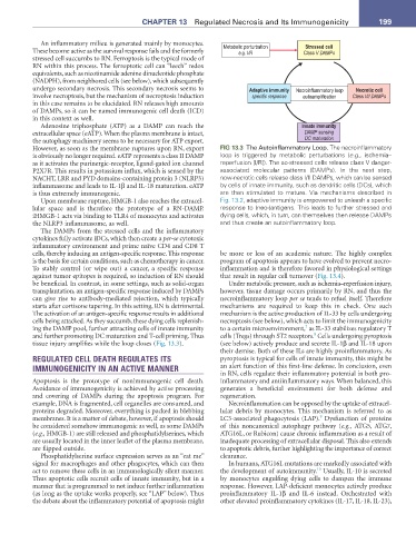
An inflammatory milieu is generated mainly by monocytes. Metabolic perturbation Stressed cell
These become active as the survival response fails and the formerly e.g. I/R Class V DAMPs
stressed cell succumbs to RN. Ferroptosis is the typical mode of
RN within this process. The ferroptotic cell can “leech” redox
equivalents, such as nicotinamide adenine dinucleotide phosphate
(NADPH), from neighbored cells (see below), which subsequently
undergo secondary necrosis. This secondary necrosis seems to Adaptive immunity Necroinflammatory loop Necrotic cell
involve necroptosis, but the mechanism of necroptosis induction specific response autoamplification Class I/II DAMPs
in this case remains to be elucidated. RN releases high amounts
of DAMPs, so it can be named immunogenic cell death (ICD)
in this context as well.
Adenosine triphosphate (ATP) as a DAMP can reach the Innate immunity
extracellular space (eATP). When the plasma membrane is intact, DAMP sensing
the autophagy machinery seems to be necessary for ATP export. DC maturation
However, as soon as the membrane ruptures upon RN, export FIG 13.3 The Autoinflammatory Loop. The necroinflammatory
is obviously no longer required. eATP represents a class II DAMP loop is triggered by metabolic perturbations (e.g., ischemia–
as it activates the purinergic-receptor, ligand-gated ion channel reperfusion [I/R]). The so-stressed cells release class V danger-
P2X7R. This results in potassium influx, which is sensed by the associated molecular patterns (DAMPs). In the next step,
NACHT, LRR and PYD domains-containing protein 3 (NLRP3) now-necrotic cells release class I/II DAMPs, which can be sensed
inflammasome and leads to IL-1β and IL-18 maturation. eATP by cells of innate immunity, such as dendritic cells (DCs), which
is thus extremely immunogenic. are then stimulated to mature. Via mechanisms described in
Upon membrane rupture, HMGB-1 also reaches the extracel- Fig. 13.2, adaptive immunity is empowered to unleash a specific
lular space and is therefore the prototype of a RN-DAMP. response to (neo-)antigens. This leads to further stressed and
iHMGB-1 acts via binding to TLR4 of monocytes and activates dying cells, which, in turn, can themselves then release DAMPs
the NLRP3 inflammasome, as well. and thus create an autoinflammatory loop.
The DAMPs from the stressed cells and the inflammatory
cytokines fully activate iDCs, which then create a per-se cytotoxic
inflammatory environment and prime naïve CD4 and CD8 T
cells, thereby inducing an antigen-specific response. This response be more or less of an academic nature. The highly complex
is the basis for certain conditions, such as chemotherapy in cancer. program of apoptosis appears to have evolved to prevent necro-
To stably control (or wipe out) a cancer, a specific response inflammation and is therefore favored in physiological settings
against tumor epitopes is required, so induction of RN should that result in regular cell turnover (Fig. 13.4).
be beneficial. In contrast, in some settings, such as solid-organ Under metabolic pressure, such as ischemia–reperfusion injury,
transplantation, an antigen-specific response induced by DAMPs however, tissue damage occurs primarily by RN, and thus the
can give rise to antibody-mediated rejection, which typically necroinflammatory loop per se tends to refuel itself. Therefore
starts after cortisone tapering. In this setting, RN is detrimental. mechanisms are required to keep this in check. One such
The activation of an antigen-specific response results in additional mechanism is the active production of IL-33 by cells undergoing
cells being attacked. As they succumb, these dying cells replenish- necroptosis (see below), which acts to limit the immunogenicity
7
ing the DAMP pool, further attracting cells of innate immunity to a certain microenvironment, as IL-33 stabilizes regulatory T
8
and further promoting DC maturation and T-cell priming. Thus cells (Tregs) through ST2 receptors. Cells undergoing pyroptosis
tissue injury amplifies while the loop closes (Fig. 13.3). (see below) actively produce and secrete IL-1β and IL-18 upon
their demise. Both of these ILs are highly proinflammatory. As
REGULATED CELL DEATH REGULATES ITS pyroptosis is typical for cells of innate immunity, this might be
IMMUNOGENICITY IN AN ACTIVE MANNER an alert function of this first-line defense. In conclusion, even
in RN, cells regulate their inflammatory potential in both pro-
Apoptosis is the prototype of nonimmunogenic cell death. inflammatory and antiinflammatory ways. When balanced, this
Avoidance of immunogenicity is achieved by active processing generates a beneficial environment for both defense and
and covering of DAMPs during the apoptosis program. For regeneration.
example, DNA is fragmented, cell organelles are consumed, and Necroinflammation can be opposed by the uptake of extracel-
proteins degraded. Moreover, everything is packed in blebbing lular debris by monocytes. This mechanism is referred to as
9
membranes. It is a matter of debate, however, if apoptosis should LC3-associated phagocytosis (LAP). Dysfunction of proteins
be considered somehow immunogenic as well, as some DAMPs of this noncanonical autophagy pathway (e.g., ATG5, ATG7,
(e.g., HMGB-1) are still released and phosphatidylserines, which ATG16L, or Rubicon) cause chronic inflammation as a result of
are usually located in the inner leaflet of the plasma membrane, inadequate processing of extracellular disposal. This also extends
are flipped outside. to apoptotic debris, further highlighting the importance of correct
Phosphatidylserine surface expression serves as an “eat me” clearance.
signal for macrophages and other phagocytes, which can then In humans, ATG16L mutations are markedly associated with
10
act to remove these cells in an immunologically silent manner. the development of autoimmunity. Usually, IL-10 is secreted
Thus apoptotic cells recruit cells of innate immunity, but in a by monocytes engulfing dying cells to dampen the immune
manner that is programmed to not induce further inflammation response. However, LAP-deficient monocytes actively produce
(as long as the uptake works properly, see “LAP” below). Thus proinflammatory IL-1β and IL-6 instead. Orchestrated with
the debate about the inflammatory potential of apoptosis might other elevated proinflammatory cytokines (IL-17, IL-18, IL-23),