
Page 221 - Clinical Immunology_ Principles and Practice ( PDFDrive )
P. 221
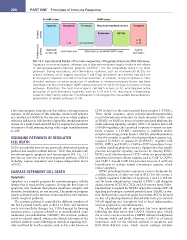
200          ParT ONE  Principles of Immune Response


                             No DAMP release                     DAMP release

                               Apoptosis  Necroptosis  Ferroptosis  Mitochondria-mediated necrosis  Pyroptosis
                          Cell death  pathway                    Parthanatos  MPT-RN


                                                                                          IL-1β
                                             IL-33  Lipid peroxidation                    IL-18
                          Proinflammatory  potency  (in vitro only)  Local inflammation  Inflammation by uncontrolled DAMP release  inflammation
                               Neglectable
                                                                                         Systemic

                       FIG 13.4  Hypothetical Model of the Immunogenicity of Regulated Necrosis (RN) Pathways.
                       Apoptosis is nonimmunogenic, whereas loss of plasma membrane integrity results in the release
                                                                 5,6
                       of damage-associated molecular patterns (DAMPs)  into the extracellular space in all other
                       pathways. During necroptosis, antiinflammatory cytokines, such as interleukin-33 (IL-33), are
                       actively released, which triggers regulatory T cell (Treg) recruitment and thereby may limit the
                       immunogenic response to a distinct microenvironment. In contrast, during ferroptosis or mito-
                       chondrial necrosis, no active production of cytokines or immunomodulatory factors has been
                       described, and the uncontrolled DAMP release accounts for the immunogenic potency of these
                       pathways. Pyroptosis, the most immunogenic cell death known so far, encompasses active
                       production of proinflammatory cytokines, such as IL-1β and IL-18, resulting in a long-lasting,
                       systemic inflammatory response. The difference in immunogenicity may explain the evolutionary
                       preservation of several pathways of RN.


        a low immunogenic stimulus can thus induce a strong immune   CD95 or Apo1) or the tumor necrosis factor receptor 1 (TNFR1).
        response. In the presence of this stimulus, common self epitopes   These death receptors, upon trimerization/hexamerization,
        are classified as DAMPs by the immune system, which explains   recruit downstream molecules via death domains (DDs), such
        why mice deficient in LAP develop a lupus-like autoinflammatory   as TRADD or FADD, to form a receptor-associated platform, the
        disease. As a result, functional LAP may be required for prevention   death-inducing signaling complex (DISC). If nuclear factor-κB
        of memory B-cell priming during solid-organ transplantation   (NF-κB)–signaling (the canonical response to tumor necrosis
        as well.                                               factor receptor 1 [TNFR1] activation) is inhibited and/or
                                                               receptor-interacting protein kinase 1 (RIPK1) polyubiquitination
        SIGNALING PATHWAYS OF REGULATED                        is lost, this complex is capable of activating initiator caspases (e.g.,
        CELL DEATH                                             caspase-8 [CASP-8] or caspase-10 [CASP-10]). Together with
                                                               RIPK1, RIPK3, and FADD, a CASP-8–cFLIP heterodimer forms
        RCD is an umbrella term for any genetically determined signaling   a cellular signaling platform named a ripoptosome that usually
                                      11
        pathway that results in cellular demise.  RCD thus includes both   prevents necroptotic signaling (see below) by cleaving RIPK1,
        nonimmunogenic apoptosis and immunogenic RN.  Fig. 13.5   RIPK3, and Cylindromatosis (CYLD) while not proteolytically
        provides an overview of the most important pathways of RCD,   activating downstream effector caspases, such as CASP-3, CASP-6,
        including  caspase-dependent  and  caspase-independent  forms   and CASP-7. Should CASP-8 be activated, however, it will form
        of RCD.                                                homodimers to activate named downstream caspases, which
                                                               induce apoptosis.
        CASPASE-DEPENDENT CELL DEATH                              RIPK1 polyubiquitination represents a major checkpoint for
                                                               a cellular decision of either survival or RCD. For this reason, it
        Apoptosis                                              is tightly regulated. Inhibitors of apoptosis 1 and 2 (cIAP1/2)
        Apoptosis is a complex program for nonimmunogenic cellular   and the linear ubiquitination complex (LUBAC) add ubiquitin
        demise that is regulated by caspases. During the first hours of   chains, whereas OTULIN, CYLD, and A20 remove them. Polyu-
        apoptosis, cells maintain their plasma membrane integrity and   biquitination is required for NEMO-dependent canonical NF-κB
        therefore, by definition, are not necrotic. The apoptotic program   signaling and mitogen-activated protein kinase (MAPK) activa-
        may be activated via the intrinsic (mitochondrial) or the extrinsic   tion. Both provide a survival signal. In contrast, RCD is licensed
        (death receptor) pathways.                             on loss of polyubiquitination. Remarkably, both pro-survival
           The intrinsic pathway is controlled by different members of   NF-κB signaling and necroptosis lead to local inflammation,
        the Bcl-2 protein family, such as Bcl-2 or BAX, and therefore   whereas apoptosis is noninflammatory.
        reacts to intracellular changes (e.g., DNA damage) by forming   The physiological role of apoptosis has been questioned
        transient pores, a process referred to as mitochondrial outer   following the observation that CASP-8 deficient mice (which
        membrane permeabilization (MOMP). The intrinsic pathway   die in utero) can be rescued on a RIPK3-deficient background
        reacts to internal stimuli, whereas the default activation of the   to  become  viable  and  fertile.  However,  CASP-8  is  of  critical
        extrinsic pathway occurs following an outside-in signaling, typi-  importance only for the extrinsic pathway of apoptosis, and
        cally mediated by death receptors, such as Fas (also known as   BAX-/BAK-deficient  mice,  which  cannot  undergo  intrinsic