
Page 223 - Clinical Immunology_ Principles and Practice ( PDFDrive )
P. 223
202 ParT ONE Principles of Immune Response
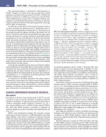
The canonical pathway is initiated by inflammasomes as Virus Bacteria/ DAMPs TNFα
NLRP3 (Chapter 3), which forms upon stimuli that include
pathogen-associated molecular patterns (PAMPs), DAMPs, and DAI TLR3/4 TNFR1
absent in melanoma-2 (AIM2) as a response to cytosolic DNA.
These inflammasomes recruit CASP-1 via adapter molecule ASC TRIF RIPK1
(apoptosis-associated speck-like protein containing a C-terminal
caspase recruitment domain) and lead to gasdermin D cleavage
and IL-1β/IL-18 maturation. RIPK3 RIPK3 pRIPK3
As both the noncanonical and canonical pathways lead to
necrotic cell death, the question about its mechanism arose. It
is clear that proinflammatory cytokines pro-IL-1β and pro-IL-18 pMLKL pMLKL pMLKL
are actively matured by caspases, resulting in the release of IL-1β FIG 13.6 Necroptosis Induction. Different triggers are known
and IL-18 from the cytosol into the extramembrane space upon to induce necroptosis, and they are sensed by specific adaptors.
necrosis execution. It is a matter of debate if, in parallel, another Viruses are sensed by DNA-dependent activator of interferon
mechanism of release of IL-1β and IL-18 exists and functions regulatory factors (DAIs), which subsequently triggers RIPK3
while the plasma membrane remains intact. In this context, polymerization by replacing RIPK1 (receptor-interacting protein
maturation is independent of gasdermins, but release is not. So kinase 1). Bacterial pathogen-associated molecular patterns
how do N-terminal gasdermin fragments kill cells and release (PAMPs) and other DAMPs are sensed by Toll-like receptors
cytokines? There is evidence that these fragments form oligomers, (TLRs) 3 and 4, which trigger RIPK3 polymerization by RHIM-
which can bind to selected negatively charged lipids, such as domain containing protein Toll–interleukin-1 receptor [TIR]–
phosphoinositide and cardiolipin. After docking to membranes, domain–containing adapter-inducing interferon-β (TRIF). The
pores are formed. If these are directly formed by gasdermins or best-described pathway is activated upon tumor necrosis factor
just mediated by them (or even artificial, see also “Necroptosis”) (TNF)-α stimulation of TNF receptor 1, which leads to RIPK1
remains a matter of debate. Phosphoinositides are restricted to phosphorylation. This phosphorylation licenses RIPK3 polymeriza-
the inner leaflet of mammalian membranes, whereas cardiolipin tion as well. As result of the active necrosome based on the
is restricted to mitochondrial and bacterial membranes. This poly-RIPK3-backbone, mixed linage kinase domain-like protein
might protect bystander cells and reduce numbers of intracellular (MLKL) is phosphorylated and triggers membrane permeabilization
bacteria before necrotic release. and CXCL1/IL-33 transcription in the nucleus.
In pyroptosis, proinflammatory factors are released. Indeed,
12
active production of systemically effective inflammatory cytokines of interferon regulatory factors (DAIs). Recently, DAI (also
during RN has been exclusively described for pyroptosis, thus known as ZBP1) has been demonstrated to mediate in utero
rendering this cell death modality the most inflammatory. A lethality of RIPK1-deficient mice by forcing RIPK3 oligomeriza-
working model for the immunogenicity of various RCD pathways tion, which places inactive RIPK1 as an inhibitor of necroptosis.
is presented in Fig. 13.4. Pyroptosis, as all other pathways or RN, The RIPK1 kinase inhibitor necrostatin-1 (Nec-1) seems to
therefore exhibits an example of ICD. stabilize this conformation and thereby inhibit necroptosis on
As expected of a cell death subroutine that causes systemic death receptor activation by not releasing RIPK1s’ inhibition to
inflammation downstream of an inflammasomal trigger, pyrop- RIPK3 oligomerization.
tosis is typically observed in macrophages during gram-negative Downstream of the active necrosome, mixed lineage kinase-like
infection and upon culture with bacterial or viral intracellular domain protein (MLKL) becomes phosphorylated by RIPK3
pathogens. Pyroptosis is thus thought to mediate the immuno- (see Fig. 13.5). After phosphorylation of MLKL, this pseudokinase
genic destruction of colonized niches. forms oligomers targeting the plasma membrane via its four-
helical bundle (4HB) motif to mediate plasma membrane rupture
CASPASE-INDEPENDENT REGULATED NECROSIS and necrotic cell death by currently undefined means. Phos-
phorylated MLKL (pMLKL) is required, but is not sufficient to
Necroptosis execute necroptosis. Whether pMLKL directly forms pores or
Necroptosis is the best characterized mode of RN. It was dis- mediates pore formation is unknown (as well as for gasdermins
covered as a type of necrotic cell death in apoptosis-resistant in pyroptosis). However, this is of great interest as pMLKL targets
cell lines. Classically, it is induced upon tumor necrosis factor-α multiple intracellular membranes, translocates to the nucleus
(TNF-α) stimulation whilst apoptosis is blocked (e.g., by a to induce CXCL1/IL-33, and is stably expressed in terminally
pan-caspase inhibitor, such as zVAD). In this context, TNFR1, differentiated cells, such as podocytes and endothelia, without
Fas, and other death receptors (described at pathways of killing these. Taken together, this might point to a still-unknown
extrinsic apoptosis, see above) transduce this signal into the cell physiological role of pMLKL.
(Fig. 13.6). Mice deficient in RIPK3 or FADD and MLKL die following
RIPK1, a key checkpoint of cellular fate, contains a unique challenge with influenza A virus, which has been demonstrated
motif next to its DD referred to as RIP homotypic interacting to depend on DAIs’ RHIM domain. Viruses also indirectly
motif (RHIM). This motif is contained in only four proteins activate necroptosis by JAK-STAT–dependent protein kinase R
within the whole mammalian proteome, and all these proteins upregulation.
are associated with the regulation of necroptosis. The necrosome, Bacterial infection is sensed via TLR3 and TLR4. This also
a higher order structure with a poly-RIPK3 backbone, is central recruits RHIM domain–containing protein TRIF, which leads
to necroptosis execution and can, next to named death receptor– to necrosome formation. Current understanding favors the idea
associated pathway, also be engaged by TRIF-binding TLR3 and that necroptosis is an evolutionary conserved program to defend
TLR4, as well as by viral sensing protein DNA-dependent activator against viruses and certain bacteria. In line with this, some viruses