
Page 386 - Clinical Immunology_ Principles and Practice ( PDFDrive )
P. 386
368 Part tHrEE Host Defenses to Infectious Agents
somatic hypermutation and affinity maturation for the selection Primary infection Recall infection
of high-affinity, antibody-producing, long-lived plasma cells, as Expansion Contraction Memory
17
well as memory B cells. At the molecular level, upregulation
of the transcription factors Blimp-1, XBP-1, and IRF-4 dictates
plasma cell formation, whereas Pax-5 expression delineates B
cells destined for GC reactions and the memory B-cell lineage.
Antibody binding to epitopes expressed by native proteins at
the surface of free virions usually blocks viral attachment or
penetration of target cells. Sometimes the consequence is viral Antigen - specific T cells
lysis (with complement proteins also involved), opsonization,
or sensitization for destruction by Fc receptor–bearing cells that
mediate antibody-dependent cellular cytotoxicity (ADCC).
Occasionally, however, Fc receptor binding of antibody-bound
virus may facilitate infection and result in more severe tissue Time
damage. This occurs in Dengue fever and may happen in some A
instances in HIV infection. The antibody involved in the protec-
tion of mucosal surfaces in humans is predominantly secretory Naive Effector 90–95% death
immunoglobulin A (IgA), but serum-derived IgG may also be T EM T CM
13
protective, particularly in such sites as the vaginal mucosa.
Both antibody isotypes act mainly to block infection of epithelial
cells, although in some instances, the antibody may transport hi hi
antigen from within the body across epithelial cells to the outside. CD62L hi CD62L lo CD62L lo lo CD62L hi
CCR7
CCR7
Mucosal antibody persists for a much shorter period compared CD69 lo CCR7 lo CCR7 lo CD69 lo
CD69
with serum antibody, which explains, in part, why immunity to T RM
mucosal pathogens is usually of much shorter duration compared CD62L lo
with immunity to systemic viral infections. CCR7 lo
CD69 hi
B
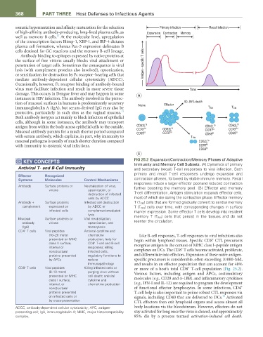
KEY CONCEPtS FIG 25.2 Expansion/Contraction/Memory Phases of Adaptive
Immunity and Memory Cell Subsets. (A) Dynamics of primary
Antiviral T- and B-Cell Immunity and secondary (recall) T-cell responses to viral infection. Both
primary and recall T-cell responses undergo expansion and
Effector recognized
Systems Molecules Control Mechanisms contraction phases, followed by stable immune memory. Recall
responses induce a larger effector pool and reduced contraction
Antibody Surface proteins or Neutralization of virus, further boosting the memory pool. (B) Effector and memory
virions opsonization, or T-cell differentiation. Antigen stimulation expands effector cells,
destruction of infected
cells by ADCC most of which die during the contraction phase. Effector memory
Antibody + Surface proteins Infected cell destruction T (T EM ) cells that are formed gradually convert to central memory
complement expressed on by ADCC or T (T CM ) cells over time, with corresponding changes in surface
infected cells complement-mediated marker expression. Some effector T cells develop into resident
lysis memory T (T RM ) cells that persist in the tissues and do not
Mucosal Surface proteins or Viral neutralization, reenter the circulation.
antibody virions opsonization, and
(IgA) transcytosis
CD4 T cells Viral peptides Antiviral cytokine and
+
(10–20 mers) chemokine Like B-cell responses, T-cell responses to viral infections also
presented on MHC production; help for begin within lymphoid tissues. Specific CD8 CTL precursors
+
+
class II surface, CD8 T-cell and B-cell recognize antigen in the context of MHC class I–peptide antigen
internal or responses; killing +
nonstructural infected cells; complexes on DCs. The CD8 T cells become activated, proliferate,
proteins presented regulatory functions to and differentiate into effectors. Expansion of these naïve antigen-
by APCs reduce specific precursors is considerable, often exceeding 10 000-fold,
immunopathology and results in an effector population that can account for 40%
+
CD8 T cells Viral peptides Killing infected cells or or more of a host’s total CD8 T-cell population (Fig. 25.2).
+
(8–10 mers) purging virus without Various factors, including antigen and APCs, costimulatory
presented on MHC cell death; antiviral
class I surface, cytokine and molecules (e.g., CD28 and 4–1BB), and inflammatory cytokines
internal, or chemokine production (e.g., IFN-I and IL-12) are required to program the development
+
nonstructural of functional effector lymphocytes. In some infections, CD4
proteins presented T-cell help is also important to prime robust CTL responses via
on infected cells or signals, including CD40 that are delivered to DCs. Activated
18
by cross-presentation CTL effectors then exit lymphoid organs and access almost all
ADCC, antibody-dependent cellular cytotoxicity; APC, antigen- body locations via the bloodstream. However, effectors do not
presenting cell; IgA, immunoglobulin A; MHC, major histocompatibility stay activated for long once the virus is cleared, and approximately
complex. 95% die by a process termed activation-induced cell death.