
Page 388 - Clinical Immunology_ Principles and Practice ( PDFDrive )
P. 388
370 Part tHrEE Host Defenses to Infectious Agents
survives the contraction phase and gradually differentiates into T RM cells can trigger enhanced early inflammation to drive local
24
a stable memory population. Upon reinfection, these memory immunity. This is in contrast to T EM cells, which continue to
cells can be rapidly activated and, by virtue of their increased migrate through nonlymphoid tissues, rather than being seques-
+
frequency, mediate more rapid clearance of the viral pathogen. tered in peripheral tissues, and also differs from the CD8 and
+
Moreover, repeated stimulation of memory cells via multiple CD4 T CM , which migrate largely through lymphoid organs (spleen
infections with the same virus, or prime-boost vaccine regimes, and lymph nodes). These differences may define the physiological
further increases the size of the antigen-specific memory T-cell raison d'être for these memory T-cell subsets, highlighting that
25
pool. Restimulation also affects the activation status and tissue measurement of memory T cells in human peripheral blood is
distribution of memory T cells, which may enhance protection a poor representation of the total-body memory T cell pool.
from viral infection in mucosal and other tissues. T RM cells can be detected in tissues by using markers, such
Experiments in humans and mice have demonstrated that as CD69 and CD103, although these are imperfect identifiers,
memory T cells are heterogeneous. Memory T cells were divided including in human tissues. T RM cells in different anatomical
into effector memory (T EM ) and central memory (T CM ) subsets, locations share a common genetic signature and require common
defined by expression of two surface molecules involved in T-cell transcription factors for their formation. Yet, these cells also
lo
24
lo
migration: CD62L and CCR7. The CD62L CCR7 T EM subset adopt unique gene expression that is imprinted by the tissue
is found primarily in nonlymphoid tissues and the spleen, whereas environment, and presumably imparts specialized functions on
hi
hi
the CD62L CCR7 T CM subset is largely present in lymph nodes T RM cells in each location. However, memory in certain peripheral
and the spleen. The current model predicts that effector T cells tissues, such as lungs, appears to wane over time, suggesting that
form the T EM subset and that these cells gradually convert to a memory T cells may not persist in sufficient numbers in this
T CM phenotype over time (Fig. 25.2B). Although the conditions site. This rationalizes a need for vaccines that induce optimal
that control the rate of this conversion are unknown, it is likely numbers of memory T cells in tissues as well as blood.
that the amounts of antigen and inflammatory signals received
during the effector phase greatly influence this. It has also been
+
shown that CD4 T-cell help is required for the generation of IMMUNE EVASION AND IMMUNITY TO CHRONIC
+
long-lived memory CD8 T cells, via interactions with DCs. 21 VIRAL INFECTIONS
Studies suggest that T CM are capable of mounting stronger
proliferative responses following reinfection. Tissue-specific Many, if not all, viruses employ immune blunting or delay tactics
homing of T EM cells permits them to enter sites of potential viral to circumvent aspects of the immune system, allowing them
27
infection, such as skin and mucosae. However, we now know time to replicate further or escape detection (Table 25.3). One
that many memory T cells found at sites of previous viral infec- such mechanism may involve killing or infecting APCs. Viruses
26
tions take up long-term residence in tissues. This includes skin, may also delay or prevent apoptosis induced by CTLs within
+
intestines, lungs, the liver, and the brain. These resident memory infected cells. Other viral evasion measures aimed at the CD8 T
T cells (T RM cells) are sequestered from the circulation and provide cell–mediated antiviral defense system inhibit antigen processing,
rapid protection against viruses, such as HSV, in skin, where thereby minimizing effector CTL induction. To escape CTL killing,
they localize with a unique dendritic morphology and undergo many viruses also downregulate the MHC molecules on the
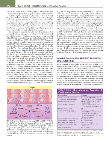
slow surveillance of the tissue (Fig. 25.3). Notably, activation of surface of infected cells. In addition, viruses may produce various
mimics or modulators/inhibitors of cytokines, chemokines, or
Epidermis
TABLE 25.3 Mechanisms and Examples of
Viral Immune Evasion
Mechanism Example
Interference with viral antigen HSV (ICP47), EBV (EBNA-1), HIV
processing and presentation (Nef, Tat), HPV (E5), CMV (UL6)
Evasion of NK cell function HIV (Nef), EBV (EBNA-1), CMV
(UL40, UL18)
+
+
CD8 T RM Basement CD8 T RM Inhibition of cell apoptosis Adenovirus (RID complex and
membrane E1B), HIV (Nef), EBV (BHRF-1)
Destruction of T cells HIV
Interference with antiviral EBV (IL-10 homologue),
+
CD4 T
CD4 T EM EM cytokines and chemokines CMV(US28 chemokine receptor
+
homologue), vaccinia virus
(IL-18-binding protein), HIV (Tat
chemokine activity)
Dermis
Inhibition of complement action HSV, pox viruses
+
+
FIG 25.3 Unique Subsets of Memory CD8 and CD4 T Cells Inhibition of DC maturation HSV, vaccinia virus
Reside Within Peripheral Tissues, at Sites of Previous Viral Frequent antigenic variation Influenza virus, HIV
Infection, and Provide Rapid Protection Against Reinfection. Infection of immune privileged Measles virus, VZV and HSV
(neurons)
site
+
Resident memory CD8 T cells (T RM ) remain localized in the Immune exhaustion HIV, HCV, HBV
epidermis in skin after herpes simplex virus (HSV) infection.
Resident memory CD4 T (T EM ) cells continue to migrate through CMV, cytomegalovirus; DC, dendritic cell; EBV, Epstein–Barr virus; HBV, hepatitis B
+
the dermal layers of skin, with access to blood and lymphoid virus; HCV, hepatitis C virus; HIV, human immunodeficiency virus; HPV, human
papillomavirus; HSV, herpes simplex virus; IL-18, interleukin-18; NK, natural killer; RID,
tissues. receptor internalization and degradation; VZV, varicella-zoster virus.