
Page 402 - Clinical Immunology_ Principles and Practice ( PDFDrive )
P. 402
CHAPtER 26 Host Defenses to Intracellular Bacteria 383
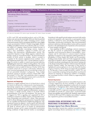
TABLE 26.3 Antibacterial Effector Mechanisms of Activated Macrophages and Corresponding
Microbial Evasion Strategies
Macrophage Effector Mechanism Microbial Evasion Strategy
Production of ROIs Uptake via complement receptors; production of ROI detoxifying molecules
(superoxide dismutase, catalase); bacterial ROI scavengers (phenolic
glycolipids, sulfatides, lipoarabinomannans)
+
Production of RNIs Inhibition of phagosome maturation via blockage of H ATP pump, indirect
effect of ROI-detoxifying molecules
Autophagy, intraphagolysosomal killing Egression into cytoplasm;
Resistant cell wall
Phagosomal acidification, phagosome–lysosome fusion Inhibition of phagosome maturation
Defensins Modification of cell wall lipid A to resist defensins
Reduced iron supply (transferrin receptor downregulation, lipocalins) Expression of microbial siderophores to increase iron uptake
Tryptophan degradation Upregulation of bacterial tryptophan synthesis
ATP, adenosine triphosphate; ROI, reactive oxygen intermediate; RNI, reactive nitrogen intermediate.
as IFN-γ and TNF, and microbial products, such as LPS, lipo- Noninfected cells engulf bacterial antigens associated with vesicles
teichoic acid, and mycobacterial lipids. RNIs exert their bactericidal produced by apoptotic cells. Apoptosis as a prerequisite for this
activity by destroying iron-/sulfur-containing reactive centers pathway is induced by many intracellular bacteria, including
of bacterial enzymes and by synergizing with ROIs to form highly salmonellae, mycobacteria, and listeriae. This cross-presentation
−
reactive peroxynitrite (ONOO ). Despite being highly effective pathway in infections with intracellular bacteria adds an essential
in killing intracellular bacteria, NO production relies on a continu- function to the physiological role of apoptosis in the maintenance
ous supply of L-arginine, which becomes limited because of of tissue integrity and growth.
competition with another macrophage enzyme, Arginase-1 Upon signaling via IFN-γ, autophagy, a process common to
(Arg-1). Arg-1 metabolizes L-arginine to produce urea and all cells for removal of dysfunctional or damaged cellular
ornithine and demonstrates antiinflammatory activity. The organelles, can be harnessed to dispose of intracellular L. mono-
competitive function of Arg-1 likely regulates collateral tissue cytogenes and M. tuberculosis in a process termed xenophagy.
damage caused by overexuberant RNIs. The final downstream Signaling via members of the immunity-related guanosine tri-
product of NOS2 activity is citrulline, which is recycled to phosphatase (GTPase) family (IRG family) and the guanylate-
L-arginine by the enzymes argininosuccinate synthase (Ass1) binding protein family, TLR2 and TLR4 engagements and the
and argininosuccinate lyase (Asl). A mouse deficient in macro- active form of vitamin D 3 all act to augment xenophagy. Formation
phage Asl activity is unable to control mycobacterial infection, of double-membrane autophagosomes that mature analogously
highlighting the importance of this recycling pathway. The central to the phagosomal pathway and fuse with lysosomes that degrade
role of NOS2 in protection against intracellular bacteria is well bacteria contained within. The importance of this process is
established in murine models of infection. Whether NOS2 plays highlighted by polymorphisms in one of the three IRG families
a similarly central role in humans is still unclear. Defensins are of genes in humans, IRGM, being associated with susceptibility
small lysosomal polypeptides that are microbicidal at basic pH to TB. Recently, a host-encoded microRNA, miRNA-155, was
and are particularly abundant in phagocytes. These include shown to potentiate xenophagy during intracellular mycobacterial
granulysin, present in granules of human natural killer (NK) infection by targeting an endogenous inhibitor of autophagy,
28
and cytolytic T (CTLs) cells, and cathelicidin, which is regulated Ras homologue enriched in brain (Rheb) by suppressing Rheb
by vitamin D in a TLR-dependent manner and is converted by expression.
cleavage to the antimicrobial peptide LL-37.
Nutrient Deprivation
Apoptosis and Autophagy Deprivation of required nutrients to intracellular bacteria is also
Apoptosis is a highly regulated form of cell death that is critical a strategy employed by the host, markedly so within infected
29
for control of cell turnover, a vital process for tissue homeostasis. macrophages. Tryptophan degradation is achieved by the enzyme
Macrophage apoptosis also constitutes a defense mechanism, indoleamine 2,3-dioxygenase (IDO), which degrades tryptophan
allowing removal of phagocytes containing intracellular bacteria to kynurenine (see Table 26.3). This reaction is induced by IFN-γ
without the need to generate significant inflammation. Apoptosis, in both MPs and IFN-γ-responsive nonprofessional phagocytes
in contrast to cellular necrosis, results in cell death without and inhibits the growth of C. psittaci and C. trachomatis inside
permeabilization of the host cell membrane. The process can human macrophages and epithelial cells. Similarly, augmentation
be triggered by TNF-α signaling and augmented by IFN-γ, of NOS2 by IFN-γ and TNF-α depletes intracellular L-arginine,
resulting in activation of cellular caspases, mitochondrial mem- also required for growth of intracellular bacteria. 20
brane permeability, and cytochrome c release. These processes
result in cellular disintegration and generation of apoptotic bodies
that are engulfed and digested by neighboring phagocytic cells. EVASION FROM, INTERFERENCE WITH, AND
Apoptosis is protective against L. monocytogenes and Salmonella RESISTANCE TO MICROBIAL KILLING
spp. and is inhibited by M. tuberculosis, which promotes necrotic
cell death of infected cells to its benefit via mitochondrial Strategies Against Toxic Effector Molecules
membrane damage and by caspase-independent mechanisms Many intracellular bacteria have exploited successful strategies
during conditions of high bacterial burden in macrophages. against macrophage effector mechanisms (see Table 26.3). One