
Page 413 - Clinical Immunology_ Principles and Practice ( PDFDrive )
P. 413
394 PARt tHREE Host Defenses to Infectious Agents
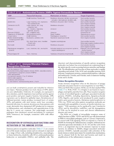
TABLE 27.2 Antimicrobial Proteins (AMPs) Against Extracellular Bacteria
AMP tissue/Cell Sources Mechanism of Action target Organisms
α-defensins Small intestines, Paneth cells Membrane disruption; inhibits complement Gram-positive bacteria
activation; chemoattracts dendritic cells Gram-negative bacteria
β-defensins Large intestines, skin, respiratory Membrane disruption; lipid II binding Gram-positive bacteria
tract epithelial cells Gram-negative bacteria
Cathelicidin (LL37) Large intestine, skin, lung, Membrane disruption Gram-positive bacteria
urogenital tract Gram-negative bacteria
RNases Skin, intestine, respiratory Unknown Gram-positive bacteria
epithelia, placenta Gram-negative bacteria
Psoriasin (S100A7) Skin, urogenital tract Unknown Escherichia coli
Calprotectin (S100A8-A9) Abscesses/neutrophils Metal Chelation Staphylococcus aureus
C-type lectins Small intestines Peptidoglycan recognition Gram-positive bacteria
Bactericidal/permeability- Neutrophils, epithelial cells Neutralizes lipopolysaccharide Gram-negative bacteria
increasing protein (BPI)
Lysozyme Skin, body fluids, tears, intestinal Degrades peptidoglycan Gram-positive bacteria
Paneth cells Some gram-negative bacteria activity
Dermcidin Sweat glands Membrane disruption Gram-positive bacteria
Gram-negative bacteria
Peptidoglycan recognition Liver, intestines, skin, neutrophils Activates bacterial two-component systems; Especially active against gram-
proteins targets peptidoglycan positive bacteria
Gram-negative bacteria
Phospholipase A2 Tears, intestines Hydrolysis of bacterial phospholipids Gram-positive bacteria
TABLE 27.3 Immune Microbial Pattern discovery and characterization of specific pattern recognition
Recognition Molecules molecules (see below) has revolutionized our understanding of
the initial specific events occurring between microbes and human
Toll-like receptors (TLRs) cells. The identification and function of these molecules is rapidly
Nucleotide-binding oligomerization domain (NOD), caterpillar proteins, expanding and include TLRs, NOD and caterpillar proteins, RNA
peptidoglycan recognition proteins (PGRPs) helicases, complement proteins, antimicrobial peptides, collectins
RNA helicases/PkR
Complement proteins: C1q, C1 inhibitor and surfactants, C-lectins, and S-lectins, such as mannose-binding
Antimicrobial peptides lectin and L-ficolin.
Collectins and surfactants
C- and S-lectins: mannose-binding lectin, L-ficolin Pattern Recognition Receptors
Innate immune recognition relies on the detection of unique
8,9
molecular structures found on microorganisms by host PRRs.
and are both constitutively present and inducible by infection TLRs and NOD-like receptors (NLRs) are the best studied PRRs
7
and injury. Humans produce two main classes of APPs: defen- (Table 27.4). TLRs (TLR1–11) (Chapter 3) are found on macro-
sins and cathelicidins. Defensins, for example, are expressed in phages, neutrophils, and other host cells. These receptors recognize
skin, intestines, and the respiratory tract and have activity against a variety of microbial ligands or pathogen-associated molecular
gram-positive and gram-negative bacteria. Interestingly, APPs patterns (PAMPs), including lipoproteins, lipopolysaccharide
expression can be altered by epithelial injury. Keratinocytes of (LPS), flagellin, and nucleic acids produced by gram-negative
inflamed psoriatic lesions produce increased levels of certain and/or gram-positive bacteria. Alterations (polymorphisms) in
APPs, and patients with such lesions rarely have secondary TLRs (e.g., TLR4) and other pattern recognition molecules are
bacterial infections. In contrast, keratinocytes from patients with associated with susceptibility or severity of specific infections (e.g.,
10
atopic dermatitis have dampened APP production in response sepsis). TLR expression can be regulated by type I interferons
to inflammation, and colonization and superinfections of the (IFNs) and by microRNAs (miRNAs), and the dysregulation
skin by S. aureus are common. Not surprisingly, successful of TLRs can be involved in acute and chronic inflammatory
pathogens have developed several mechanisms to counteract diseases and cancer. 11
APPs. Gonococci, for example, counteract APPs with efflux The NLRs are a family of intracellular receptors, some of
8
pumps. which function as PRRs. NOD1 and NOD2 are well characterized
as PRRs to extracellular pathogens, such as S. flexneri. Importantly,
in concert with TLR signaling, NLR can respond to a variety of
RECOGNITION OF EXTRACELLULAR BACTERIA AND PAMPs by forming the inflammasome complex. Inflammasome
ACTIVATION OF THE IMMUNE SYSTEM activation generates interleukin-18 (IL-18) and the active form
of IL-1, an important step in the immune response to many
Immune pattern recognition molecules (Table 27.3) are a major bacteria. In addition to PAMPs, danger-associated molecular
arm of the innate immune system and are released or expressed patterns (DAMPs), typically associated with activation of the
by a range of host cells, including lymphocytes, macrophages, innate immune system upon injury by noninfectious mechanisms,
or tissue histiocytes, dendritic cells (DCs), polymorphonuclear can be released both by the host and bacteria and result in
leukocytes or neutrophils (PMNs), and epithelial cells. The amplification of the inflammatory response.