
Page 415 - Clinical Immunology_ Principles and Practice ( PDFDrive )
P. 415
396 PARt tHREE Host Defenses to Infectious Agents
deficiency, to name a few). Bacterial infections associated with
phagocytic dysfunction are described elsewhere (Chapter 22).
Innate Lymphoid Cells
Innate lymphoid cells (ILCs), a heterogeneous group of cells of
the innate immune system, have lymphoid morphology but lack
the capacity for rearrangement of the antigen receptors, a cardinal
18
feature of the cells of the adaptive immune system. Conventional
natural killer (NK) cells, better known for generating inflam-
matory cytokines and cytotoxic activity against malignant cells
and cells infected by viruses, seem to have a role in the defense
against bacterial pathogens. Murine study data suggest that
conventional NK-cell activation by lung macrophages is protective
19
against S. aureus pneumonia. A subset of ILCs, known as NK-like
cells, produces IL-22 and has been found in mucosal sites, where
these ILCs appear to have a protective activity against bacterial
FIG 27.2 Bacterial Phagocytosis at Mucosal Surfaces. Transmis- pathogens. IL-22 derived from these cells modulates AMP expres-
sion electron micrograph of phagocyte engulfing Neisseria sion by epithelial cells. NK-like cells have minimal cytotoxic
meningitidis at a human respiratory epithelial mucosal surface activity and are not strong producers of IFN-γ, core characteristics
41
(×19 000). of conventional NK cells. 20
Lymphocytes
Carbohydrate Th1 cells are characterized by IFN-γ and function to activate
capsule
macrophages to phagocyte and kill pathogens. While this
Antiphagocytic mechanism of pathogen elimination is primarily directed against
structures pathogens with a predominant intracellular lifecycle, Th1 cells
are relevant for typical extracellular bacteria as pneumococcus
21
and S. aureus. The neutrophilic response to extracellular bacteria
Secreted Pilus is primarily coordinated by Th17 cells. Animal models have
22
bacterial Leukocidin
products C5a peptidase suggested that the Th17 response is central for protection against
that lyse a wide variety of gram-positive and gram-negative bacteria. For
phagocytes or example, Th17 response has been shown to induce nasopharyngeal
impede chemotaxis clearance of Pneumococcus in both animal models and in children.
Differentiation toward the Th17 subtype appears to be favored
by strong antigenic signals and broad activation of pathogen
recognition receptors. IL-17 and IL-22, the signature interleukins
(ILs) of the Th17 response, promote AMP secretion by epithelial
cells, neutrophil migration, and epithelial integrity. The increased
susceptibility of subjects with Job’s syndrome to S. aureus infec-
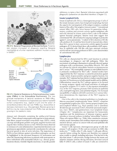
FIG 27.3 Bacterial Resistance to Polymorphonuclear Leuko- tions demonstrates the importance of the Th17 response in
cytes (PMNs) in the Extracellular Environment. The two humans.
principal mechanisms of bacterial resistance to PMN killing. One in five cells in the intestinal epithelium is a lymphocyte.
These consist of resistance to phagocytosis as a result of bacterial Mucosa-associated lymphoreticular tissue (MALT) comprises
surface components (e.g., capsule or pili) and the action of intraepithelial lymphocytes (IELs), lamina propria lymphocytes,
extracellular proteins that can lyse PMNs (e.g., leukocidins) or and lymphoid follicles (e.g., Peyer patches) and is sometimes
decrease chemotaxis (e.g., C5a peptidase). Bacteria growing in divided into gut-associated (GALT), bronchial-associated (respira-
biofilms may be more protected from PMNs than are bacteria tory tract) (BALT), and genitourinary tract lymphoid tissues
growing in the planktonic state. (Chapter 20). Lymphocytes are important for homeostatic
23
regulation and the maintenance of immune response against
elastase) and chromatin containing the antibacterial histone microbes at mucosal surfaces, including “extracellular” bacteria.
17
H2A. These released compounds work together to form extracel- These cells express PRRs (e.g., TLRs), have constitutive cytotoxic
lular fibers, termed neutrophil extracellular traps (NETs), which activity, secrete chemokines and cytokines important in regulation
can trap and kill gram-positive and gram-negative bacteria and and host defense, and act in concert with mucosal epithelial cells
degrade their virulence factors as well. NETs have been observed and exocrine glands.
in instances of acute inflammation (experimental dysentery and Innate T cells represent a heterogeneous group of cells that
spontaneous appendicitis) and provide a mechanism for reducing possess T-cell receptors (TCRs) and are restricted to MHC-like
bacterial spread at sites of acute infection. The importance of molecules. 24,25 Unlike other T cells, innate T cells gain effector
PMNs in host defense against extracellular pathogens can best capacity before exiting the thymus and therefore can respond
be highlighted by the increased frequency of bacteremias and more readily to stimuli, including infections. This has led to the
other life-threatening infections in patients with neutropenia or idea that innate T cells provide a bridge between the innate and
those individuals with neutrophil deficits (e.g., chronic granu- adaptive immune systems during infections. Invariant natural
lomatous disease, Chediak-Higashi syndrome, or specific granule killer T (iNKT) cells, a subset of innate T cells, are restricted to