
Page 414 - Clinical Immunology_ Principles and Practice ( PDFDrive )
P. 414
CHAPtER 27 Host Defenses to Extracellular Bacteria 395
4
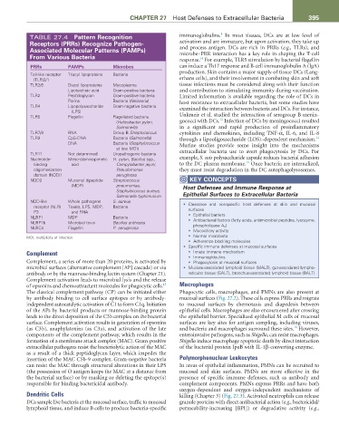
TABLE 27.4 Pattern Recognition immunoglobulins. In most tissues, DCs are at low level of
Receptors (PRRs) Recognize Pathogen- activation and are immature, but upon activation, they take up
Associated Molecular Patterns (PAMPs) and process antigen. DCs are rich in PRRs (e.g., TLRs), and
microbe–PRR interaction has a key role in shaping the T-cell
From Various Bacteria response. For example, TLR5 stimulation by bacterial flagellin
14
PRRs PAMPs Microbes can induce a Th17 response and B-cell immunoglobulin A (IgA)
Toll-like receptor Triacyl lipoproteins Bacteria production. Skin contains a major supply of tissue DCs (Lang-
(TLR)2/1 erhans cells), and their involvement in combating skin and soft
TLR2/6 Diacyl lipoproteins Mycoplasma tissue infections must be considered along with their function
Lipoteichoic acid Gram-positive bacteria and contribution to stimulating immunity during vaccination.
TLR2 Peptidoglycan Gram-positive bacteria Limited information is available regarding the role of DCs in
Porins Bacteria (Neisseria) host resistance to extracellular bacteria, but some studies have
TLR4 Lipopolysaccharide Gram-negative bacteria examined the interaction between bacteria and DCs. For instance,
(LPS)
TLR5 Flagellin Flagellated bacteria Unkmeir et al. studied the interaction of serogroup B menin-
12
(Helicobacter pylori, gococci with DCs. Infection of DCs by meningococci resulted
Salmonella) in a significant and rapid production of proinflammatory
TLR7/8 RNA Group B Streptococcus cytokines and chemokines, including TNF-α, IL-6, and IL-8
TLR9 CpG-DNA Bacteria (Salmonella) through a lipooligosaccharide (LOS)–dependent mechanism.
12
DNA Bacteria (Staphylococcus Murine studies provide some insight into the mechanisms
at low MOI)
TLR11 Not determined Uropathogenic bacteria extracellular bacteria use to avert phagocytosis by DCs. For
Nucleotide- Meso-diaminopimelic H. pylori, Bacillus spp., example, S. suis polysaccharide capsule reduces bacterial adhesion
15
binding acid Campylobacter jejuni, to the DC plasma membrane. Once bacteria are internalized,
oligomerization Pseudomonas they must resist degradation in the DC autophagolysosomes.
domain (NOD)1 aeruginosa
NOD2 Muramyl dipeptide Streptococcus KEY CONCEPtS
(MDP) pneumoniae, Host Defenses and Immune Response at
Staphylococcus aureus,
Salmonella typhimurium Epithelial Surfaces to Extracellular Bacteria
NOD-like Whole pathogens S. aureus • Clearance and nonspecific host defenses at skin and mucosal
receptor (NLR) Toxins, LPS, MDP, Bacteria
P3 and RNA surfaces
NLRP1 MDP Bacteria • Epithelial barriers
NLRP1b Microbial toxin Bacillus anthracis • Antibacterial factors (fatty acids, antimicrobial peptides, lysozyme,
phospholipase A 2 )
NLRC4 Flagellin P. aeruginosa
• Mucociliary activity
• Normal microbiota
MOI, multiplicity of infection.
• Adherence blocking molecules
• Specific immune defenses at mucosal surfaces
Complement • Innate immune mechanism
• Immunoglobulins
Complement, a series of more than 20 proteins, is activated by • Phagocytosis at mucosal surfaces
microbial surfaces (alternative complement [AP] cascade) or via • Mucosa-associated lymphoid tissue (MALT), gut-associated lympho-
antibody or by the mannose-binding lectin system (Chapter 21). reticular tissue (GALT), bronchus-associated lymphoid tissue (BALT)
Complement activation leads to microbial lysis and the release
13
of opsonins and chemoattractant molecules for phagocytic cells. Macrophages
The classical complement pathway (CP) can be initiated either Phagocytic cells, macrophages, and PMNs are also present at
by antibody binding to cell surface epitopes or by antibody- mucosal surfaces (Fig. 27.2). These cells express PRRs and migrate
independent autocatalytic activation of C1 to form C1q. Initiation to mucosal surfaces by chemotaxis and diapedesis between
of the APs by bacterial products or mannose-binding protein epithelial cells. Macrophages are also encountered after crossing
leads to the direct deposition of the C3b complex on the bacterial the epithelial barrier. Specialized epithelial M cells of mucosal
surface. Complement activation results in generation of opsonins surfaces are key sites for antigen sampling, including viruses,
16
(as C3b), anaphylatoxins (as C3a), and activation of the late and bacteria and macrophages surround these sites. However,
components of the complement pathway, which results in the enteroinvasive pathogens, such as Shigella, can resist macrophages.
formation of a membrane attack complex (MAC). Gram-positive Shigella induce macrophage apoptotic death by direct interaction
extracellular pathogens resist the bacteriolytic action of the MAC of the bacterial protein IpaB with IL-1β-converting enzyme.
as a result of a thick peptidoglycan layer, which impedes the
insertion of the MAC C5b-9 complex. Gram-negative bacteria Polymorphonuclear Leukocytes
can resist the MAC through structural alterations in their LPS In areas of epithelial inflammation, PMNs can be recruited to
(the possession of O antigen keeps the MAC at a distance from mucosal and skin surfaces. PMNs are more effective in the
the bacterial surface) or by masking or deleting the epitope(s) presence of specific immune defenses, such as antibody and
responsible for binding bactericidal antibody. complement components. PMNs express PRRs and have both
oxygen-dependent and oxygen-independent mechanisms of
Dendritic Cells killing (Chapter 3) (Fig. 27.3). Activated neutrophils can release
DCs sample live bacteria at the mucosal surface, traffic to mucosal granule proteins with direct antibacterial action (e.g., bactericidal/
lymphoid tissue, and induce B cells to produce bacteria-specific permeability-increasing [BPI]) or degradative activity (e.g.,