
Page 417 - Clinical Immunology_ Principles and Practice ( PDFDrive )
P. 417
398 PARt tHREE Host Defenses to Infectious Agents
antigens. Antigen mimicry can lead to autoantibodies, as in the LPS, PG monomers, DNA repeats
case of rheumatic fever and glomerulonephritis after S. pyogenes Lipoproteins, Teichoic acid
infection. Antigen mimicry can also dampen the immune response Microbial toxins, other microbial components
to bacterial antigens, as in the case of serogroup B Meningococcus. (Superantigens)
Other microbial surface structures, such as the pili of the gonococ-
cus, can “stiff-arm” neutrophils, keeping them at a distance. A Pattern recognition receptors (e.g., TLRs)
number of pyogenic bacteria (e.g., S. aureus) secrete leukocidins,
which lyse phagocytes. Other pathogens (e.g., group A strepto-
cocci) inhibit chemotaxis of neutrophils through the elaboration Cytokine stimulation
of enzymes (e.g., C5a peptidase) that proteolytically cleave
chemotactic signals. Some bacteria possess mechanisms to prevent Coagulopathy TNF-a Complement activation
30
opsonization by changing surface antigens. Many bacteria form Kinin stimulation IL-1 C5a
biofilms, which shield these microorganisms from host defense Prostaglandins INF-γ C3a
34
molecules and antibiotics. Leukocytes that invade S. aureus Leukotrienes IL-6, IL-8 Leukocyte chemotaxis
biofilms exhibit impaired phagocytosis and decreased ability to PAF IL-10 Inflammation
kill bacteria. In addition, biofilm matrices can protect bacteria
from antibody-mediated phagocytosis. Fibrin deposition Nitric oxide
As previously noted, many “extracellular” bacteria have an DIC
intracellular component to their lifecycle. The intracellular
environment provides protection from proteins of the comple- Generalized endothelial damage
ment system, Igs, and nonspecific barriers to infection present Vascular leak
28
in the epithelia. The entry of bacteria into epithelial cells provides Tissue edema
access to nutrients and protection from host defenses, allows Vasodilation
protected multiplication, and leads to shedding of organisms Leukocyte activation
Bleeding
back to the mucosal surface, to facilitate transmission and further Temperature dysregulation (e.g. fever)
spread of the infection on the epithelium. Attachment can also
initiate epithelial cell apoptosis or toxin-mediated cell death and Tachycardia, hyperventilation
lead to the breakdown of the epithelial barrier. Hypotension (↑CO, ↓SVR)
Pallor, peripheral vasoconstriction
Cutaneous signs
HOST RISK FACTORS FOR LOCAL AND SYSTEMIC Multiorgan failure
INVASION BY EXTRACELLULAR PATHOGENS (ARDS, renal failure)
Altered mental status
Bacteria that breach mucosal and skin barriers and reach sub- Shock
Death
mucosal tissues of sites, such as pulmonary alveoli or the middle
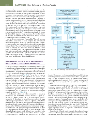
ear and/or the bloodstream, induce immune responses, including FIG 27.5 Inflammatory cascade initiated during sepsis.
cytokine release, phagocytosis, complement activation, antibody
release or production, and other local or systemic induction of
the inflammatory cascade (Fig. 27.5). The survival of bacteria invasive bloodstream meningococcal and gonococcal infections,
following colonization of the epithelium and access to the indicating an important role for insertion of the complement
bloodstream depends on the integrity of the host immune MAC in the bactericidal activity of human serum against
response (including variability caused by genetic polymorphisms) pathogenic Neisseria. In adults, 10–20% of invasive meningococcal
and on the ability of the bacteria to resist this host immune disease has been associated with a defect in the complement
response. Host factors that increase the risk for the development system.
of systemic disease as a result of extracellular bacteria include In infants, antibacterial activity wanes as levels of passively
polymorphisms in innate immune mechanisms, the absence of transferred maternal antibody fall. This waning of antibody is
bactericidal or opsonizing antibodies, deficiencies in the comple- correlated with the highest incidence of several “extracellular”
ment pathways, and an absence of or reduction in neutrophil pyogenic bacterial diseases (caused by S. pneumoniae, N. men-
function or levels (see Table 27.1). ingitidis, H. influenzae type b) in young children. During child-
Complement deficiencies, either congenital or acquired, hood and adolescence, levels of bactericidal antibodies rise and
increase the risk for invasive bacterial diseases (Chapter 21). rates of these diseases decline. Specific antibodies are acquired
Because C3 plays a critical role in the complement cascade, through carriage and through cross-reacting epitopes on other
congenital C3 deficiency or conditions that reduce C3 (e.g., commensal species. For example, cross-reactive antibodies to N.
systemic lupus erythematosus, cirrhosis, nephritis, C3 nephritic meningitidis are acquired by colonization with commensal Neis-
factor) increase the risk for invasive disease due to pyogenic seria spp. (e.g., Neisseria lactamica) and unrelated bacteria (e.g.,
bacteria, such as S. pneumoniae and N. meningitidis. Mannose- Enterococcus faecium, Bacillus pumilus, and E. coli). The lack of
binding lectin (MBL) is a plasma opsonin that initiates comple- bactericidal antibodies against a strain recently acquired in the
ment activation. MBL gene polymorphisms are found in children upper respiratory tract is an important risk factor for invasive
with meningococcal and pneumococcal sepsis. Properdin defi- meningococcal disease.
ciency, leading to defective AP killing, is also associated with In addition to defects in innate immunity, Igs, and complement
severe and recurrent meningococcal infections. Terminal comple- deficiencies, human genetic polymorphisms are associated with
ment deficiencies (C5–C8) are also associated with recurrent an increased risk or severity of bacterial diseases. For example,