
Page 49 - Clinical Immunology_ Principles and Practice ( PDFDrive )
P. 49
CHaPter 2 Organization of the Immune System 35
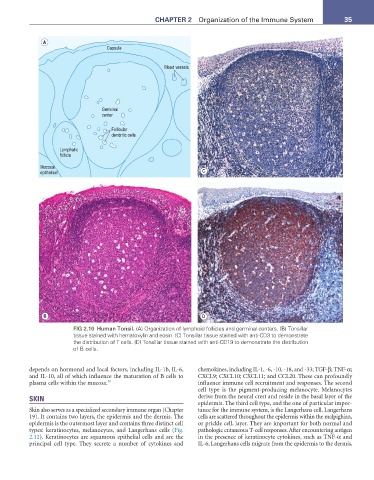
A
Capsule
Blood vessels
Germinal
center
Follicular
dendritic cells
Lymphatic
follicle
Mucosal
epithelium C
B D
FIG 2.10 Human Tonsil. (A) Organization of lymphoid follicles and germinal centers. (B) Tonsillar
tissue stained with hematoxylin and eosin. (C) Tonsillar tissue stained with anti-CD3 to demonstrate
the distribution of T cells. (D) Tonsillar tissue stained with anti-CD19 to demonstrate the distribution
of B cells.
depends on hormonal and local factors, including IL-1b, IL-6, chemokines, including IL-1, -6, -10, -18, and -33; TGF-β; TNF-α;
and IL-10, all of which influence the maturation of B cells to CXCL9; CXCL10; CXCL11; and CCL20. These can profoundly
plasma cells within the mucosa. 56 influence immune cell recruitment and responses. The second
cell type is the pigment-producing melanocyte. Melanocytes
SKIN derive from the neural crest and reside in the basal layer of the
epidermis. The third cell type, and the one of particular impor-
Skin also serves as a specialized secondary immune organ (Chapter tance for the immune system, is the Langerhans cell. Langerhans
19). It contains two layers, the epidermis and the dermis. The cells are scattered throughout the epidermis within the malpighian,
epidermis is the outermost layer and contains three distinct cell or prickle cell, layer. They are important for both normal and
types: keratinocytes, melanocytes, and Langerhans cells (Fig. pathologic cutaneous T-cell responses. After encountering antigen
2.12). Keratinocytes are squamous epithelial cells and are the in the presence of keratinocyte cytokines, such as TNF-α and
principal cell type. They secrete a number of cytokines and IL-6, Langerhans cells migrate from the epidermis to the dermis,