
Page 604 - Clinical Immunology_ Principles and Practice ( PDFDrive )
P. 604
582 ParT FIvE Allergic Diseases
which suppress inflammation. Additional therapies for moderate production of matrix metalloproteinases (MMPs) and other
to severe disease include theophylline and the phosphodiesterase enzymes that degrade elastin (elastases), including MMP2, MMP9,
inhibitor roflumilast. During exacerbations of COPD, which MMP12, and neutrophil elastase (NE). The activity of most of
become increasingly frequent with advancing disease and are these enzymes is controlled by endogenous inhibitors, such as
often caused by bacterial and viral infections, tailored use of tissue inhibitors of metalloproteinases (TIMPs) and A1AT, the
antibiotics is frequently helpful. For severe disease with resting principal antagonist of elastases. Cigarette smoke exposure is
hypoxemia, continuous nasal oxygen can both extend life and sufficient to enhance elastase secretion from macrophages and
relieve dyspnea. Surgical interventions, such as lung volume neutrophils and to reduce the activity of elastase inhibitors. In
reduction surgery and lung transplantation, are reserved for addition to elastin, MMP12 specifically inhibits A1AT, leading
advanced disease. to unopposed NE activity. These biochemical events are critically
The immunological basis of COPD differs markedly from important to the expression of emphysema because elastin is an
asthma: smoking-related lung inflammation appears to be the important MMP that is partly responsible for both the structural
fundamental underlying cause of both airway obstruction and integrity of the lung and its stretchiness. In genetically susceptible
lung destruction (Fig. 41.9). In contrast to asthma, where Th2 individuals, elastin becomes the antigenic target of pathological
cells and other allergic effector cells are involved, the major Th1 and Th17 cells. Emphysema, therefore, is the clinical expres-
immune effector cells in COPD are Th1 and Th17 cells. Together sion of the autoimmune-based loss of lung integrity resulting
with macrophages and neutrophils, these innate and adaptive from enhanced expression of antielastin immunity that promotes
immune cells coordinate lung destruction in COPD by enhancing secretion of elastases. 42
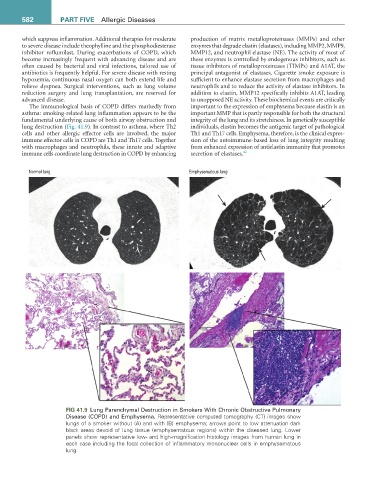
Normal lung Emphysematous lung
FIG 41.9 Lung Parenchymal Destruction in Smokers With Chronic Obstructive Pulmonary
Disease (COPD) and Emphysema. Representative computed tomography (CT) images show
lungs of a smoker without (A) and with (B) emphysema; arrows point to low attenuation dark
black areas devoid of lung tissue (emphysematous regions) within the diseased lung. Lower
panels show representative low- and high-magnification histology images from human lung in
each case including the focal collection of inflammatory mononuclear cells in emphysematous
lung.