
Page 600 - Clinical Immunology_ Principles and Practice ( PDFDrive )
P. 600
578 ParT FIvE Allergic Diseases
TABLE 41.2 Chemoattractants
'HFOLQH )(9 ,QFUHDVH 5 Linked to the recruitment of allergic receptor
Inflammatory Cells
Chemokine
CCL1 CCR8
CCL11 CCR3
)ROORZ XS KRXUV CCL17, CCL22 CCR4
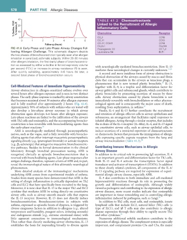
FIG 41.6 Early-Phase and Late-Phase Airway Changes Fol- CX 3 CL1 CX 3 CR1
Prostaglandin D2
CR T H 2
lowing Allergen Challenge. This schematic diagram depicts Leukotriene B4 BLT1
the two phases of bronchoconstriction typically seen after allergen
inhalation in sensitized, asthmatic subjects. Within 20–30 minutes
after allergen inhalation, the first (early) phase of bronchoconstric-
tion as assessed by either a decline in forced expiratory volume
per second (FEV 1) or increase in airway resistance (R) is seen. with neurologically mediated bronchoconstriction. How IL-13
11,25
After quickly subsiding, approximately 4–6 hours (h) later, a mediates these neurological changes is currently unknown.
second (late) phase of bronchoconstriction occurs. A second and more insidious form of airway obstruction is
physical obstruction of the airways caused by mucus and fibrin
clots that can accumulate in the airways as tenacious plugs, a
26
phenomenon that is now termed plastic bronchitis. IL-13,
Cell-Mediated Features of Immediate Hypersensitivity together with IL-9, is a trophic and differentiation factor for
Airway obstruction in allergen-sensitized asthma evolves over airway goblet cells and submucosal glands, which contribute to
several hours after allergen exposure and is seen in two distinct plastic bronchitis by promoting secretion of mucus by these
phases. The early-phase response is marked by airway constriction cells. Airway obstruction caused by plastic bronchitis is not
that becomes maximal about 30 minutes after allergen exposure immediately reversible with bronchodilators or other pharma-
and is fully resolved after approximately 2 hours (Fig. 41.6). cological agents and is consequently the major cause of death,
Approximately 50% of subjects with asthma who are tested will resulting from asphyxiation, in asthma. 27
also develop a late-phase airway response in which airway Finally, IL-4 and IL-13 further coordinate the recruitment
obstruction again develops 4–6 hours after allergen exposure. and retention of allergic effector cells to airway epithelium and
Late-phase reactions are linked to the infiltration of the airways submucosa, an arrangement that facilitates rapid responses to
with Th2 cells and eosinophils, and the accompanying broncho- inhaled allergens. Acting through a similar receptor, that includes
constriction is less reversible with bronchodilating agents than the α chain of the IL-4 receptor (IL-4Rα), IL-4, and IL-13 signal
the early-phase reactions. 24 on constitutive airway cells, such as airway epithelial cells, to
AHR is neurologically mediated through parasympathetic induce secretion of a restricted repertoire of chemoattractants
nerves, such as the vagus, and is fully reversible with broncho- or chemotactic factors that promote the immigration of allergic
dilating agents that either interrupt muscarinic parasympathetic cells expressing specific cognate receptors from the lung and
signaling directly (e.g., ipratropium bromide) or activate receptors airway microcirculation (Table 41.2). 28
(e.g., β 2 -adrenergic) that antagonize muscarinic bronchoconstric-
tive pathways. Besides its formal demonstration in the clinical Contributing Immune Mechanisms in Allergic
laboratory through bronchial provocation testing, AHR is Airway Disease
recognized clinically as episodic bronchoconstriction that is In addition to its critical role in promoting IgE secretion, IL-4
reversed with bronchodilating agents. Late-phase responses after is an important growth and differentiation factor for Th2 cells.
antigen challenge, therefore, represent a form of AHR and, in part, Both IL-13 and IL-4 activate the transcription factor signal
reveal the immunological nature of this ultimately neurological transducer and activator of transcription 6 (STAT6). In addition
phenomenon. to these closely related cytokines, all components of the IL-4/
More detailed analysis of the immunological mechanisms IL-13 signaling pathway are required for expression of experi-
underlying AHR comes from experimental models of asthma. mental allergic airway disease, especially AHR.
Studies from many species have demonstrated that AHR in the IL-5 also contributes to both immediate and cell-mediated
setting of allergic inflammation is critically dependent on Th2 hypersensitivity reactions through its role in promoting the
cells and ILC2 that have specifically been recruited to the lung. growth and differentiation of eosinophils. Although widely
Moreover, it is now clear that IL-13 is the major Th2 and ILC2 viewed as pathogenic and contributing to the expression of allergic
cytokine that mediates AHR by acting directly on constitutive airway diseases, more recent studies indicate that eosinophils
airway cells, such as airway smooth muscle cells that express are important in tissue remodeling and in controlling allergic
the IL-13 receptor. However, IL-13 does not directly induce inflammation induced by pathogens, such as fungi. 29
bronchoconstriction. Bronchoconstriction in subjects with In addition to Th2 cells, mast cells, and eosinophils, innate
asthma, expressed as episodic bouts of dyspnea, is triggered by lymphoid cells that include ILC2, natural killer (NK) cells (a
diverse exogenous factors in addition to allergens (e.g., altered type of ILC1), and γδ T cells may also contribute to allergic
temperature and humidity, pungent odors, irritating aerosols) disease expression through their ability to rapidly secrete Th2
and endogenous stimuli (e.g., extreme emotional states) with and other cytokines. 25
little apparent connection to immunological mechanisms. Numerous additional soluble mediators contribute to the
Thus rather than directly mediating airway obstruction, IL-13 expression of allergic disease. The complement system is especially
establishes the basis for responding broadly to diverse agents important, and complement proteins C3a and C5a, the major